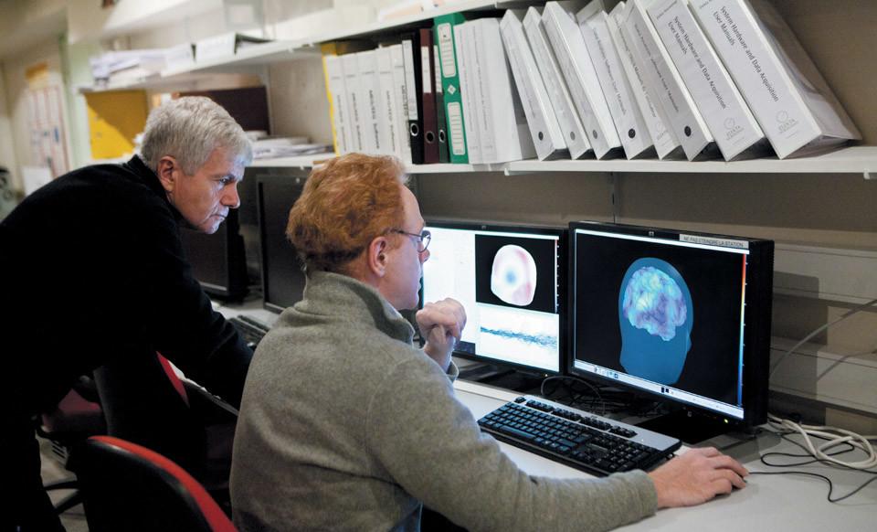
The brain's total white matter volume (WMV) increases rapidly from the second trimester to early childhood, peaking at age 28.7 and then accelerating its decline after age 50. (Visual China/Photo)
Years ago, Jakob Seidlitz, a neuroscientist at the University of Pennsylvania, took his 15-month-old son to a pediatrician for a check-up, and the doctor simply used height and weight charts to check and thought his child was developing normally. However, as a neuroscientist, Saidlitz knows that the brain is the most important organ for cognitive things and the world, so at least there should be some way to detect the brain to know whether a person's brain is normal from birth to old age, whether there are any changes, so as to provide a scientific basis not only for improving health, but also for improving the brain's cognition. Shockingly, however, pediatricians lack reliable biological information about the development of children's brain organs.
Based on the idea of accurately detecting and evaluating the brain, Saidlitz and his team later did a huge and arduous study, collecting human magnetic resonance imaging (MRI) for study. MRI can provide information such as the shape and volume of the human brain from the fetus to the time of death, so that the dynamic conditions of the human brain with time can be intuitively observed and understood. The findings were published in the April 6, 2022 issue of the journal Nature.
In total, the team collected 123894 MRI scans of 101457 people, ranging from fetuses at 16 weeks of pregnancy to 100-year-olds. MRI scans are diverse not only in age, but also in types, including the brains of healthy normal people, the brains of people with various diseases (such as Alzheimer's), and neurocognitive differences (such as autism spectrum disorder). The researchers used statistical models to extract information from images and ensure that scan results were directly comparable regardless of the type of MRI machine used. From this, they created the most comprehensive chart of human brain development to date. These charts visually show how the human brain grows rapidly early in life and then slowly shrinks with age.
Using modeling recommended by the World Health Organization, Saideritz et al. created brain maps of human lifespans based on a generalized additive model (GAMLSS) of position, proportion, and shape. GAMLSS and related statistical frameworks have previously been applied to open datasets to assess brain function throughout a person's life cycle.
At the same time, the team integrated the GAMLSS model into the subjects' brains with four main tissue volumes, including total cortical gray matter volume (GMV), total white matter volume (WMV), total subcortical gray matter volume (sGMV), and total ventricular cerebrospinal fluid volume (ventricle or cerebrospinal fluid) to assess brain changes with age.
The function of gray matter and white matter of the brain is not the same, gray matter is the nerve center, plays a role in innervation, including polio, can also control some low-level non-conditioned reflexes, white matter is mainly responsible for conduction, such as spinal white matter mainly conducts the excitement of the brain and polio. Anatomically, gray matter and white matter have different distributions. In the brain and cerebellum and brainstem, the outer part is gray matter and the interior is white matter. However, in the spinal cord and bulbar, the outer part is mainly white matter, and the interior is gray matter.
The study found that the volume of these 4 main tissues of the brain varies with the time of development.
The total cortical gray matter volume (GMV) of the brain begins with a strong increase in the second trimester, peaking at age 5.9 and subsequently declining nearly linearly. Total white matter volume (WMV) increases rapidly from the second trimester to the early childhood, peaking at age 28.7 and then accelerating decline after age 50.
Compared with the total cortical gray matter volume and the total white matter volume, the total subcortical gray matter volume (sGMV, which controls body function and basic behavior) showed an intermediate growth pattern, peaking at age 14.4 years. Both the total volume of white matter (WMV) and the total volume of subcortical gray matter (sGMV) peaks were consistent with previous neuroimaging and autopsy reports. Cerebrospinal fluid (CSF) shows a growth trend before the age of 2 years, stabilizes before the age of 30, then grows slowly and linearly, and grows exponentially at the age of 60.
These data are clearly better than those observed in previous studies because data on earlier fetuses were obtained, while previous studies only started in infancy.
Using GAMLSS assessment, the researchers quantified the evolution of variability among subjects. Total cortex gray matter volume (GMV) developmental growth in the brain peaks in the 4th year of birth; total subcortical gray matter volume (sGMV) developmental growth peaks in late puberty; changes in total white matter volume (WMV) peak in the 40th year of life; cerebrospinal fluid (CSF) mutates the most at the end of a person's lifespan.
Based on these changes, it can be known that the earliest peak of brain development is at age 4 (total cortical gray matter volume), and continues into puberty (total subcortical gray matter volume), and until mid-term age 40 years (total white matter volume), of course, if you count cerebrospinal fluid, brain development is present at the end of life. Therefore, the overall evaluation is that aging in some parts of the brain begins at the age of 4, but some parts continue to grow and maintain a balance that is compatible with the physiological functions of the person throughout their lives.
Sedelitz et al. also used GAMLSS modeling to measure the development of the average cortical thickness, total surface area, and volume of 34 cortical regions of the whole brain. The results showed that the total area of the human cerebral cortex is closely related to the total volume (TCV) throughout the life cycle, and both indicators peak at age 11-12 years. However, cortical thickness peaks at age 1.7 years, consistent with previous findings, where cortical thickness increases during perinatal period and decreases later in development.
The study also showed significant regional differences in peaks in brain and nervous system development. Total cortical gray matter volume (GMV) peaks at age 5.9 years, but the age at which gray matter volume peaks in 34 cortical regions varies greatly, at ages 2-10 years, respectively. The primary sensory region of the brain reaches the earliest peak volume and declines faster after the peak; the frontotemporal joint cortical region reaches the volume peak later and declines slower after the peak; the volume peak of the ventral-caudal region cortex with earlier maturity decreases faster after peak volume, and the volume peak of the later mature dorsal-beaked cortex declines more slowly.
The results of age-related changes in the development of different parts of the brain suggest that cognition based on brain development presents a "basic perceptual-joint cortex" gradient, which has been closely related to multiple aspects of brain structure and function. In addition, the study also provides clues for diagnosing some neuropsychiatric disorders.
Alzheimer's disease can lead to neurodegeneration and loss of brain tissue, and people with this disease have less brain volume than their peers. By comparing the brain volume of patients with their peers, signs of neurodegeneration can be found, which, combined with other indicators, can be diagnosed and treated.
Of course, the results of this study are not perfect. First, most of the MRI data collected in the study came from North America and Europe, mainly white, college students, urban and wealthy people, while there were 3 data in South America and only 1 data in Africa, and the latter two accounted for about 1% of all MRI data used in the study. Therefore, the coverage of the data is not too comprehensive.
In addition, the study was only an MRI study and did not cover neurotransmitters, neural circuits, and blood supply that better reflect brain function.
But in any case, this diagram of brain development provides an important foundation for our understanding of the development of the human brain. Just like the normal values of some physiological tests in people, such as blood routine, urine routine, etc., brain development charts may provide some normal values for brain development.
Zhang Tiankan