Immunotherapy has become the fourth treatment for tumors after surgery, chemotherapy and radiation therapy. Different from conventional treatments that directly target tumor cells, immunotherapy kills tumor cells by regulating and activating the human immune system and relying on autoimmune functions. Among them, immune checkpoint suppression therapy represented by anti-CTLA-4 and PD-1 monoclonal antibodies has become a hot spot in current tumor treatment by lifting tumor immune tolerance, enhancing CD8+ T cell function and thereby eliminating tumor cells[1].
In recent years, researchers have found that microorganisms and their metabolites have an unimaginable impact on human health, especially tumor treatment. A growing body of research suggests that the gut microbiota modulates the efficacy of immunotherapy [2].
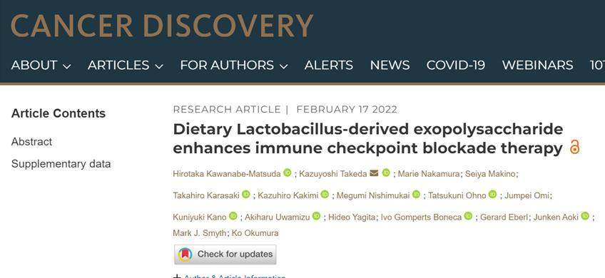
Recently, the research team led by professors at Shuntendo University in Japan found that Lactobacillus dei Bulgarian subspecies OLL1073R-1, which is common in yogurt, can secrete extracellular polysaccharides, bind to LPA receptors on the surface of CD8+ T cells through Gro3P groups, induce CCR6 expression, and enhance the anti-cancer effect of immune checkpoint inhibitors on tumors expressing CCL20. The findings[3] were published in the top international medical journal Cancer Discovery.

Screenshot of the home page of the official website paper
CD8+ T cells are the main effector cells of tumor immunity, in direct contact with tumor cells, producing effector molecules such as perforin and γ interferon, apoptosis and scavenging tumor cells. Therefore, promoting the entry of CD8+ T cells into tumor tissue is the key to achieving tumor immunotherapy [4].
The migration and infiltration of T cells is mediated by the binding of chemokines to chemokines receptors on the cell surface. Microbes in the gut, through metabolites, recruit T cells to migrate from the gut to tumors, enhancing the anti-tumor immune response [5].
Lactobacillus bulgaricus is a probiotic necessary for yogurt fermentation. Previous studies have found that Lactobacillus bulgaricus reduces the risk of infection with the common cold and influenza [6], enhances NK cell activity in mice, and induces splenocytes to produce γ interferons by producing extracellular polysaccharides[7].
So, can Lactobacillus bulgaricus also chemotactic T cells, thereby affecting immunotherapy for tumors?
First, the researchers obtained extracellular polysaccharides secreted by Lactobacillus bulgaricus and, after oral feeding to normal mice, detected the expression of T cell chemokine receptors in the intestinal pool lymph nodes of mice. The results showed that the expression level of the chemokine receptor CCR6 was significantly upregulated in CD8+ T cells. In vitro, the researchers observed the same experimental results: with Lactobacillus bulgaricus extracellular polysaccharides stimulated in the spleen or cells of peripheral blood origin, CCR6 was also specifically highly expressed in CD8+ T cells. Extracellular polysaccharides of Lactobacillus bulgaricus induce CD8+ T cells to express the chemokine receptor CCR6.
Subsequently, the researchers analyzed the relationship between CCR6 expression levels and tumor prognosis and found that in patients such as colorectal cancer, high expression of CCR6 was significantly associated with prolonged overall survival. Since the chemokine receptor CCR6 has only one ligand, the chemokine CCL20, the researchers also measured the expression of CCL20 in tumors and found that the expression of CCL20 was significantly elevated in colorectal cancer tissue. These results suggest that CCR6-expressing CD8+ T cells infiltrating CCL20-expressing tumors may indicate a good prognosis.
After oral administration of Lactobacillus bulgaricus extracellular polysaccharides (EPS-R1) in mice, Figure A: The expression level of the chemokine receptor CCR6 in CD8+ T cells in the intestinal pool lymph nodes (PP) was significantly upregulated; Figure B: Flow cytometry analysis confirmed that CCR6 expression in CD8+ T cells was upregulated; Fig. C: Survival analysis showed that the high expression of CCR6 was significantly correlated with the prolongation of overall survival of cancers such as colorectal cancer (CRAD); Figures D and E: When in vitro extracellular polysaccharides were stimulated in splenocytes, The expression ratio of CCR6 is significantly selectively increased in CD8+ T cells.
To verify the relationship between CCR6+CD8+ T cells and CCL20 in tumor tissue, the researchers conducted animal experiments.
Given that CCL20 is highly expressed in colorectal cancer and low in melanoma, the researchers constructed colorectal cancer and melanoma mice, respectively, as tumor models with different levels of expression of CCL20. In tumor mice treated with oral extracellular polysaccharides, the proportion of CD8+ T cells expressing CCR6 in colorectal cancer tissue increases with the prolongation of treatment time. This suggests that extracellular polysaccharide-induced CCR6+CD8+ T cells can selectively infiltrate into tumor tissue expressing CCL20.
Next, the researchers explored the effects of extracellular polysaccharides on immune checkpoint suppression therapy, using extracellular polysaccharides to treat colorectal cancer or melanoma mice, respectively, while immune checkpoint inhibitors were given treatment. Results The colorectal tumor body with high expression of CCL20 was significantly reduced, while the melanoma body with low expression of CCL20 had no change; after the colorectal cancer was neutralized by antibodies and CCL20, the tumor body was no longer reduced, and after the melanoma was overexpressed with CCL20, the tumor body was significantly reduced. These results suggest that extracellular polysaccharides rely on CCL20, enhancing the antitumor effects of immune checkpoint inhibitors.
Colorectal cancer (Colon26) or melanoma (B16F10) mice were treated with extracellular polysaccharides, respectively, while immune checkpoint inhibitors (aCTLA-4 mAb, aPD-1 mAb) were given. Figure G: Colorectal cancer body with high expression of CCL20 significantly reduced, Figure H: Melanoma body with low expression of CCL20 has no change, Figure I will: After melanoma overexpresses CCL20, the tumor body is significantly reduced, Figure J: Colorectal cancer is neutralized by antibody and CCL20, the tumor body is no longer shrinking.
However, how does the extracellular polysaccharide of Lactobacillus bulgaricus enhance the antitumor effect of immune checkpoint inhibitors?
In vitro, extracellular polysaccharides treat tumor cells without inducing cell death or inhibiting cell proliferation, suggesting that extracellular polysaccharides do not act directly on tumor cells to enhance anti-tumor immune responses.
In the tumor microenvironment, CD8+ T cells can produce cytokines such as γ interferon to improve the killing ability of tumor cells. So the researchers knocked out the γ interferon gene and looked at the effects of extracellular polysaccharides on tumors.
After knocking out the γ interferon gene in colorectal cancer mice, the extracellular polysaccharides were fed, which no longer had an anti-tumor enhancing effect on immune checkpoint inhibitors.
Transcriptome sequencing also found that colorectal cancer treated with extracellular polysaccharides treated with immune checkpoint inhibitors also upregulated transcriptional levels of interferon-γ interferons, and interferon-producing genes such as CXCR3.
These results show that the extracellular polysaccharide-induced CCR6+CD8+ T cells have an enhancing effect on the antitumor effect of immune checkpoint inhibitors through γ interferon mediated.
Figure A: In vitro knockout of γ interferon does not affect the induction of extracellular polysaccharides to produce CCR6+CD8+ T cells, Figure B: In vivo knockout of γ interferon causes the loss of the anti-tumor enhancement effect of extracellular polysaccharides on immune checkpoint inhibitors, Figure C: Extracellular polysaccharides combined with immune checkpoint inhibitors in the treatment of colorectal cancer, γ interferon and CXCR3 and other interferon production related genes transcriptional levels upregulated.
The presence of group 3-phosphoglycerol (Gro3P) in the extracellular polysaccharides of Lactobacillus bulgaricus has been found to be immunomodulatory. Based on this, the researchers speculate that Gro3P is a functional group of extracellular polysaccharides that exert anti-tumor-enhancing effects.
The researchers fed the extracellular polysaccharides removed from Gro3P orally to mice and found that the CCR6+ population did not increase in the CD8+ T cells in the intestinal pool lymph nodes of mice, and also lost the enhancing effect on the anti-tumor effect of immune checkpoint inhibitors. This confirms that Gro3P is key to the antineoplastic effects of extracellular polysaccharides enhancing immune checkpoint inhibitors.
Gro3P binds to hemolytic phosphatidic acid (LPA) receptors, while CD8+ T cells express LPA receptors. Therefore, the researchers again speculate that the LPA receptor of CD8+ T cells mediates the induction of extracellular polysaccharides to CCR6 expression.
The researchers conducted in vitro experiments, first using the LPA receptor agonist RP-1 to treat CD8+ T cells, and found that CCR6 expression was upregulated, while knocking out the LPA receptor of CD8+ T cells, the extracellular polysaccharide could not upregulate the expression of CCR6. This confirms that LPA receptors are necessary for CD8+ T cells to express CCR6.
Figure C: Extracellular polysaccharides and LPA are present in Gro3P groups, Figure E: Mass spectrometry to detect the presence of Gro3P groups in extracellular polysaccharides, Figure I: LPA receptor agonist RP-1 can upregulate CD8+ T cells to express CCR6, Figure J: Knock out the LPA receptor (Lpar2) of CD8+ T cells, extracellular polysaccharides cannot upregulate the expression of CCR6.
In summary, Lactobacillus Bulgaricus relies on the Extracellular Polysaccharide Gro3P group, binds to the LPA receptor on the surface of CD8+ T cells, induces chemokine receptor CCR6 expression, promotes CCR6+ CD8+ T cells infiltration into tumors expressing the chemokine CCL20, produces γ interferon, maintains T cell function, and enhances the anti-cancer effect of immune checkpoint inhibitors.
Previous studies have found that yogurt intake is associated with a reduced risk of colorectal cancer [9]. Now, Professor Takeda's team has found that Lactobacillus bulgaricus used in yogurt fermentation can secrete extracellular polysaccharides and enhance the anti-cancer effect of immune checkpoint inhibitors. However, the results and conclusions of this study are based solely on laboratory animal mice. However, compared with rodents, human genes are more diverse, contact with microbes are more diverse, in humans, the cross-dialogue between the microbiome and host is much more complex than in mice, so for "yogurt anti-cancer", it needs to be carefully viewed and further verified.
bibliography:
[1] Brunner-Weinzierl MC, Rudd CE. CTLA-4 and PD-1 Control of T-Cell Motility and Migration: Implications for Tumor Immunotherapy. Front Immunol. 2018;9:2737. Published 2018 Nov 27. doi:10.3389/fimmu.2018.02737
[2] Iida N, Dzutsev A, Stewart CA, et al. Commensal bacteria control cancer response to therapy by modulating the tumor microenvironment. Science. 2013;342(6161):967-970. doi:10.1126/science.1240527
[3] Kawanabe-Matsuda H, Takeda K, Nakamura M, et al. Dietary Lactobacillus-derived exopolysaccharide enhances immune checkpoint blockade therapy [published online ahead of print, 2022 Feb 17]. Cancer Discov. 2022;candisc.0929.2021. doi:10.1158/2159-8290.CD-21-0929
[4] Chen DS, Mellman I. Oncology meets immunology: the cancer-immunity cycle. Immunity. 2013;39(1):1-10. doi:10.1016/j.immuni.2013.07.012
[5] Klaenhammer TR, Kleerebezem M, Kopp MV, Rescigno M. The impact of probiotics and prebiotics on the immune system. Nat Rev Immunol. 2012;12(10):728-734. doi:10.1038/nri3312
[6] Guillemard E, Tondu F, Lacoin F, Schrezenmeir J. Consumption of a fermented dairy product containing the probiotic Lactobacillus casei DN-114001 reduces the duration of respiratory infections in the elderly in a randomised controlled trial. Br J Nutr. 2010;103(1):58-68. doi:10.1017/S0007114509991395
[7] Makino S, Sato A, Goto A, et al. Enhanced natural killer cell activation by exopolysaccharides derived from yogurt fermented with Lactobacillus delbrueckii ssp. bulgaricus OLL1073R-1. J Dairy Sci. 2016;99(2):915-923. doi:10.3168/jds.2015-10376
[8] Kitazawa H, Harata T, Uemura J, Saito T, Kaneko T, Itoh T. Phosphate group requirement for mitogenic activation of lymphocytes by an extracellular phosphopolysaccharide from Lactobacillus delbrueckii ssp. bulgaricus. Int J Food Microbiol. 1998;40(3):169-175. doi:10.1016/s0168-1605(98)00030-0
[9] Zhang K, Dai H, Liang W, Zhang L, Deng Z. Fermented dairy foods intake and risk of cancer. Int J Cancer. 2019;144(9):2099-2108. doi:10.1002/ijc.31959
Responsible editor 丨Ying Yuyan