The base of the skull is bounded between the base of the cranial cavity and the top of the orbit, nasopharyngeal cavity, sieve sinuses, sphenoid sinuses, and subtemporal fossa. The base of the skull is the site where many blood vessels, nerves, and special receptors are concentrated, and its anatomy is extremely complex, and there are many lesions in this area. In the daily scan will often encounter some related lesions of the skull base, many technicians do not know how to scan, this issue of the MRI scan of the skull base to do a simple combing, for your reference only.
The base of the skull is one of the most complex areas of human anatomy, and it has a large number of lesions. Many times we do not know how to scan, mainly because we are not familiar with the anatomy, do not know enough about clinical needs and pathological changes, and do not have a comprehensive understanding of the MR sequence. Familiarity with anatomy is the basis for doing a good MRI, first look at the general structure of the base of the skull.
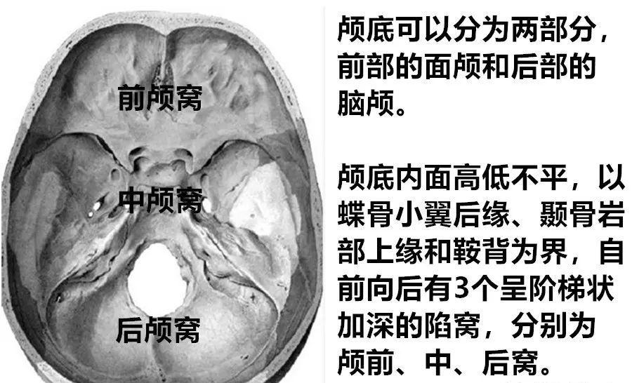
The base of the skull can be divided into two parts, the front of the skull and the posterior cranial head. There are three depressions in the base of the skull, that is, three fossa, anterior fossa, middle fossa and posterior fossa, each of which contains the corresponding tissue structure of the human body.
The anterior fossa is the shallowest and most superior of the three fossa, located above the nose and eye cavity, and the anterior fossa is equipped with an anterior and lower part of the frontal lobe. Lesions such as meningiomas, eye tumors, nasal tumors, and some neuropathic tumors are mainly involved.
Anterior skull base
FOV: 180-240mm, layer thickness/pitch 3-4mm/0.3-0.4mm
Transverse axis bit T1WI
Transverse position T2WI
The use of water grease separation is more cost-effective
Transverse position T2WI grease pressing
Transverse position DWI (optional)
Non-EPI methods are recommended
Crown T1WI or T2WI
Sagittal T2WI
intensifier
0.1mmol/kg或0.2ml/kg
Dynamic enhancement (optional)
Transverse, sagittal, coronal and/or isotropic 3D T1WI
Grease pressing is used in at least two directions
Anterior skull base scan is based on transverse axis position + coronal position, supplemented by sagittal position.
CSF leakage is suspected and a thin layer should be added to sweep the T2 image.
The use of water lipid separation T1WI can obtain better fat pressing effect.
To observe the neural structure, a 3D thin-layer scan is performed.
The middle cranial fossa is located in the center of the base of the skull and mainly involves lesions such as saddle mass, pituitary tumors, cavernous sinus lesions, and meningiomas.
Sellarica, cavernous sinuses
FOV: 180-240mm, layer thickness/spacing: 3-4mm/0.3-0.4mm
Transverse axis bit T1WI
Transverse position T2WI
The use of water grease separation is more cost-effective
Transverse position T2WI grease pressing
Transverse position DWI (optional)
Non-EPI methods are recommended
Coronal T1WI
Horizontal axis bit MRA
Doubts CCF must sweep
intensifier
0.1mmol/kg或0.2ml/kg
Dynamic enhancement (optional)
Transverse, coronal position
Grease pressing is required in both directions
Saddle pituitary gland
FOV: 160-200mm, layer thickness/spacing: 2-3mm/0.2-0.3mm
Transverse position T2WI (optional)
Assess whole-brain condition
Coronal T1WI
Coronary T2WI
Sagittal T1WI
Sagittal T2WI
When flat sweep + enhanced, it can be selected for sweeping
intensifier
Dynamic enhancements
Non-microadenomas may be scanned
Sagittal, coronal position
Grease pressing is used in at least one direction
Transverse and/or isotropic 3D T1WI
Microadenomas may be scanned
Lesions in the saddle area are dominated by coronal + sagittal positions, supplemented by transverse axial positions.
When pituitary cystic lesions are suspected, the addition of T2 FLAIR is more conducive to its differential diagnosis.
Pituitary microadenoma contrast medium uses half-dose + dynamic.
The use of water lipid separation T1WI can obtain better fat pressing effect.
The posterior fossa is the last and deepest of the three fossa. It mainly involves auditory/trigeminal neuromas, nasopharyngeal tumors, brainstem and cerebellar lesions.
Base of the posterior skull
FOV: 180-240mm, layer thickness/pitch 3-4mm/0.3-0.4mm
Transverse axis bit T1WI
Transverse position T2WI
The use of water grease separation is more cost-effective
Transverse position T2WI grease pressing
Transverse position DWI (optional)
Non-EPI methods are recommended
Crown T1WI or T2WI
Sagittal T1WI
intensifier
0.1mmol/kg或0.2ml/kg
Dynamic enhancement (optional)
Transverse, sagittal, coronal and/or isotropic 3D T1WI
Grease pressing is used in at least two directions
The sagittal position is the main orientation for assessing skull base depression, posterior fossa malformation, cerebrospinal fluid circulation, etc., and the use of isotropic 3D T1WI sequences is more conducive to the measurement and evaluation of related parameters.
To observe the neural structure, a 3D thin-layer scan is performed.
The use of water lipid separation T1WI can obtain better fat pressing effect.
The above weight comparison sequence can be achieved using 2D, 3D sequence or synthetic MRI imaging technology, and its scanning sequence can be adjusted according to the needs.
It's not so much how the base of the skull should be scanned, but rather the base of the skull can be subdivided into which scan sites. The above is only a general protocol for skull base scanning, for reference only. In actual scans, a more precise and personalized scan protocol can be developed based on the source of the lesion and the clinical needs of the patient, such as:
Eye MRI scan!
Ear MRI scan!
Pituitary MRI scan!
The general principle of grasping is the combination mode of multi-directional + multi-sequence comparison, which mainly shows the best orientation for the lesion, and the scanning method of fat pressure + non-fat pressure.

The combined mode of multi-sequence comparison highlights the greatest advantage of MRI.
As one of the most complex areas of human anatomy, the skull base contains numerous blood vessels, air chambers, fluids, bone and other tissues and structures that are easy to interfere with MRI imaging. The MRI image of the base of the skull obtained in the clinical scan is easy to produce corresponding artifacts, and some corresponding adjustments should be made according to the actual situation during the actual scan.
As one of the most complex areas of human anatomy, the base of the skull, especially during lipid compression scanning, should pay attention to the choice of sequence and method of fat pressing.
The base of the skull contains a large number of blood vessels, air chambers, fluids, bone and other tissues and structures that are easy to interfere with MRI imaging, and attention should be paid to the selection of enhancement sequences.
MRI signal as long as it comes from water and fat, anything has two sides, too much will cover up the lesion, too little will lead to insufficient contrast, in the actual scan should pay attention to the application of fat pressure and water pressure, starting from clinical needs, grasp the focus of observation, master a certain degree.
Not using lipid press can save scanning time and obtain images with a high signal-to-noise ratio, while retaining fat signals can form a natural contrast with some tissues, but high-signal fats will mask a lot of information;
The use of lipid pressure will prolong the scanning time, and the resulting image will have lower signal and noise, but some signals will be better displayed, which is more conducive to the identification of certain lesions.
The same is true of pressurized water. Whether to use fat press and pressurized water, according to the specific situation of reasonable choice.
Lipid compression scan may be used for in-fat-rich tissues or sites in enhanced scans of the base of the skull, but lipid-rich areas may be used for fat-rich areas.
Question about whether the base of the skull needs to be dynamic. In the case of unspecified diagnosis, of course, dynamic is definitely dynamic, and the dynamic enhancement time should be ≥ 5min. Dynamics can shift the focus of your observations from the display of anatomical structures to quantitative analysis, which has risen to another level for diagnosis, and it can also provide more and more accurate imaging information for clinical diagnosis and treatment.
The dynamic enhancement time should be ≥5min, and when doing the TIC curve, attention should be paid to avoiding tissues and areas that are easy to interfere with the curve, such as fluids and blood vessels.
The main problems that affect the quality of MRI images on the base of the skull are magnetic sensitivity effects and vascular beat artifacts, and the focus you need to address is this.
Tumor lesion DWI is essential, such as DWI sequences using a non-EPI approach to obtain better image quality. Above, EPI DWI on the top and non-EPI DWI on the bottom.
Especially after enhancement, the treatment of vascular beat artifacts to achieve excellent image quality is key.
bibliography:
Hu Yingying, Li Lei, Zhang Wei, Wu Xiaoling." Step-by-step" combined with sketches to illustrate the anatomy of the skull base[J].Journal of Anatomy,2021,44(6):549-552.DOI:10.3969/j.issn.1001-1633.2021.06.022.
YANG Zhenghan, FENG Feng, WANG Xiaoying. Magnetic Resonance Imaging Technical Guide[M]. People's Military Medical Publishing House, 2007.
ZHANG Zongrui. Specification for MRI scans of the inner ear and base of the skull. Beijing Tongren Hospital.2021.
CAI Hanshou. An overview of skull base anatomy and its skull base tumor imaging[C].Proceedings of the 9th Radiology Conference of Guangdong Medical Association.2007:140-141.
END
The following article is from the House of Magnetic Resonance and written by MR LiaoY
【Copyright Notice】This platform is a public welfare learning platform, reprinted for the purpose of transmitting more learning information, and has indicated the author and source, such as teachers who do not want to be disseminated can contact us to delete