MRS is currently the only functional imaging technology that can non-invasively detect chemical substances in living tissue, although the current software and hardware development and technical means of MRI have been very advanced and mature, but the application of MRS in human metabolite detection is still subject to many limitations and deficiencies. To achieve excellent spectral line quality, many aspects need to be paid attention to, such as MRI field strength, selected sequence parameters, the degree of fit of the subject, and the techniques related to the operation. This issue mainly shares common problems in MRS imaging and some personal experience summaries, for reference only.
The application of MRS is much earlier than the current clinical use of MRI tomography, but the earliest MRS is mainly used for the detection of industrial chemical substances, with the development of software and hardware and the innovation of technical means, MRS has been gradually applied to the analysis of medical human metabolites, MRS can identify abnormal signals that can not be found in conventional MRI scans.
The results obtained by MRS showed that it was not a common tomographic anatomy image in our clinic, but a metabolite component information displayed in the form of spectral lines, and the interpretation of its spectral lines was more complex than that of tomographic anatomy images.
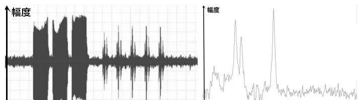
The amplitude/time information obtained by the MRI device is obtained by the Fourier transform, and its metabolites can be distinguished in the frequency domain (figure above △ , schematic diagram).
MRS imaging can detect changes and levels of corresponding metabolites caused by pathological and biochemical changes in tissues in the human body, and reflect the pathological changes in the characteristics of the disease through the information it provides.
MRS imaging in the human body is not only H1, but also P31, C13, Na23 and so on. Because the amount and class of hydrogen-containing substances in the human body are extremely rich, hydrogen protons are often selected for MRS imaging in the clinic, and we often say that MRS imaging is hydrogen atom spectroscopy.
The highest possible field strength (≥1.5 T) is recommended for MRS imaging in clinical scans.
In conventional MRI imaging, the chemical displacement phenomenon will be mitigated as much as possible, thereby alleviating the associated artifacts it brings to the image, but MRS is precisely to show different metabolites and concentrations in the human body with the help of accurate chemical displacement between different substances. The higher the field strength, the more pronounced the chemical displacement, the greater the difference between the different metabolites, and the easier it is to distinguish them in the resulting spectral lines.
The metabolites in the human body are minimal compared to the water and fat in the human body, so it is necessary to use lipid pressure and water suppression to eliminate the effect of its extremely high signal intensity on the weak metabolite signal, and use high field strength to ensure the signal-to-noise ratio of the spectral line.
The human body produces a very large number of metabolites in the metabolic process, but because of the difference in concentration between them, our current MRI imaging methods and means cannot show them all.
In the actual MRS scan should consider the feasibility and credibility of the problem, not all sites and all lesions are suitable for MRS imaging, in order to obtain the ideal spectral line, generally meet the following conditions:
There is a certain chemical displacement between the metabolites being examined, which is the basis.
Metabolites have a certain concentration, that is, the lesion cannot be too small.
Sufficient field strength can distinguish them.
Sufficient magnetic field uniformity guarantees its spectral line quality.
Relatively little lipid and water content is found in the imaging tissue or area.
In imaging voxels, try to avoid tissue structures with magnetic sensitivity effects, such as bone plates, qi sinuses, blood flow, cerebrospinal fluid, metals, necrosis, calcification, fat, and bleeding.
In THE CASE, instead of clicking directly to start the scan, it is recommended to perform a pre-scan first, obtain the relevant parameters through the pre-scan, and evaluate whether it has MRS imaging conditions before performing a formal MRS scan.
Pre-scan obtains reference information on magnetic field uniformity (line width).
Pre-scan for water suppression reference information.
On the obtained MRS line, the position of each metabolite on the line does not change with the change of field strength (ppm is independent of B0), but is fixed, and the change is the corresponding peak height and area below the line of the metabolite.

The ordinate is the signal strength.
The abscissa is a dimensionless ppm.
The reference 0 is on the right side of the coordinates, and the proton spectrum of tetramethylsilane is often used as the internal standard reference (there may be differences between different manufacturers, specific reference model operation manual).
The peak level of a single metabolite in the spectral line does not have corresponding diagnostic significance, but needs to be compared between different metabolites to have reference value.
As a semi-quantitative analysis of MRS, it lacks specific and uniform numerical standards, and the corresponding results cannot be used as the only basis for diagnosing a disease.
The alignment of the baseline is the key to obtaining an accurate area under the peak.
How to measure the quality of a spectral line, suitable for diagnosis!
The uniformity of the magnetic field (line width), depending on the narrowness of the peaks.
Signal-to-noise ratio, look at the peak height is not high.
Whether the water suppression is thorough, see if the peak dry does not interfere.
Baseline stability, look at the peaks and feet are uneven.
Choline and creatine are shown to see if they are well separated.
Even if the scan fails, be able to pinpoint the cause and adjust the scan program.
Controlling every link in MRS imaging is key, otherwise the images obtained by spending a few minutes scanning are not enough for diagnosis, then you have to scan again.
Strict braking of the person being examined.
Proper voxels. Too small a voxel can cause low signal-to-noise and unstable spectral lines.
The final line response obtained by mrs scans is a synthesis of information about all metabolites in the entire voxel. If the voxels are set too large, the normal tissues and abnormal tissue metabolites in the voxels will be averaged between each other, which will cause a partial volumetric effect, and it is not appropriate to < 10 *10 *10 mm. Similarly, if the lesion is too small, its partial volumetric effect will be obvious, and the obtained spectral line does not have reliable diagnostic value for the diagnosis of the lesion, so the diagnostic value of MRS for small lesions is extremely limited.
Precise positioning.
The use of pure (no tilt angle) transverse/sagittal/crown position is more convenient for MRS positioning, and it should be observed whether the positioned voxel avoids the organization and structure of the interfering spectral line in three directions; whether the added saturation band covers the positioned voxel.
Use saturation bands wisely.
Saturation bands are not the more the better, only placed in the easy to interfere with the signal area, such as adding a saturation band to bone and fat-rich areas to inhibit the corresponding signal.
Although enhanced images are more conducive to precise localization of lesions, contrast media may change the shape of the spectrum, so it is recommended that MRS should be performed before the contrast medium injection.
The injection of contrast agent will change the local magnetic field uniformity, resulting in an increase in the width of the spectral line, a decrease in the signal-to-noise ratio, an increase in artifacts, etc., which will affect the interpretation of the spectral line.
After the injection of contrast media will change the peak height, sub-peak area and other related parameters of some metabolites of the spectrum, although some literature shows that the injection of contrast medium has little effect on its results (more is the intracranial barrier with blood-brain barrier), but there are also literature that show that for the breast, prostate and other parts, some of the contrast medium will not only affect the quality of the spectrum, but also affect the corresponding results.
While the current literature suggests that contrast agents affect the quality of the spectral lines more, further research remains to be done on how much influence the final result is.
Rationally select scan sequences, TE values, etc. according to clinical needs.
The use of saturation bands is one of the most effective measures to improve the uniformity of the magnetic field in the imaging area. When performing MRS scanning, the saturation band should be fully utilized to improve the quality of the spectral lines, but the saturation band should not be too much, and a narrow 2-4 strips should often be used, which can be placed at a certain angle in the area prone to interference.
During MRS imaging, the system adds saturation bands parallel to the voxels by default around the voxels.
Monosomy or polyselectrons!
Monosomy
Polytels
Easy to achieve homogenization, short imaging time
It is not easy to achieve homogenization and the imaging time is longer
Voxels are larger and the signal-to-noise is higher
The signal-to-noise ratio is not as good as the MRS of monosomy
It is more conducive to the display of short T2 substances and trace substances
Imaging of larger areas is more favorable
Only a single spectral line can be obtained, not compared
Multiple spectral lines can be obtained for easy comparison
Monosomy positioning should avoid interfering with the organization and structure of the spectral line.
When multi-voxel localization, the range should include lesions, lesions surrounding the lesion, and some normal tissues for easy comparison.
STEAM (excitation echo use) or PRESS (point resolution)!
STEAM is mainly used for short-echo MRS, which has a good water suppression effect, but the signal-to-noise ratio is not as good as PRESS, and it is more sensitive to motion.
PRESS can use long and short echo MRIs, and its signal-to-noise ratio is equivalent to 2 times that of STEAM, and it is more widely used in clinical scans.
Short TE or long TE!
Short TE
Long TE
The signal strength is high, but the baseline stability is poor
The signal strength is not high, but the baseline is stable
There are many metabolites visible in the spectral lines, providing more information, but it is more difficult to quantify
Trace metabolites cannot be displayed, and there are few metabolites visible in the spectral line, which is intuitive and concise
May show lower and short T2 metabolites
Short T2 metabolites can not be displayed, which is conducive to the display of long T2 substances
Detection of specific substances, such as Lip, Glx, mI, etc
bibliography:
YANG Chun,XU Kai,ZHENG Junnian,XU Qian,MA Hong,RONG Yutao. A preliminary study on the effect of Gd-DTPA on prostate 3T hydrogen proton spectroscopy[J].Chinese Journal of Computer Imaging for Medicine,2011,17(4):332-334.DOI:10.3969/j.issn.1006-5741.2011.04.010.
Expert Consensus on MR Examination and Diagnosis[J].Chinese Journal of Radiology,2021,55(10):1008-1023.DOI:10.3760/cma.j.cn112149-20210609-00548.
YU Tonggang,DAI Jiazhong,QIAN Huijun,FENG Xiaoyuan. Analysis of influencing factors of brain MRS examination[J].Journal of Clinical Radiology,2010,29(6):716-720.
ZHANG Yingkui,LI Li,LI Jinfeng. Principle and application of magnetic resonance imaging system[M]. Peking University Medical Press, 2021.
ZHAO Bin,CAI Shifeng,YU Taifei,PENG Hongjuan,LI Huihua. Value of MR hydrogen proton spectroscopy in the application of breast mass and analysis of technical interference factors[J].Chinese Journal of Radiology,2006,40(3):281-284.DOI:10.3760/j.issn:1005-1201.2006.03.015.
YANG Zhenghan, FENG Feng, WANG Xiaoying. Magnetic Resonance Imaging Technical Guide[M]. People's Military Medical Publishing House, 2007.
JIN Erhu, JIANG Tao, ZHANG Hui. Introduction to Clinical Applications of Magnetic Resonance Imaging (2nd Edition)[J]. Chinese Journal of Clinicians, 2015(4).
B.Kastler. In-depth understanding of MRI basics[M]. People's Military Medical Publishing House, 2012.
Shen Guiquan,Qu Xueling,Gao Bo,Yu Hui,Wei Yuqing,Li Dongfang,Shi Qianhua,Xiang Zhihua. Analysis of the effect of magnetic resonance paramagnetic contrast media (Gd-DTPA) on 1H-MRS[J].Journal of Practical Radiology,2008,24(8):1119-1121.DOI:10.3969/j.issn.1002-1671.2008.08.034.
ZHANG Kai,LI Chuanfu,LIU Ying,CONG Peixin,ZHENG Jinyong,FENG Dechao,MENG Xiangshui. Effect of contrast media on MR spectroscopy of 3T hydrogen proton[J].Chinese Journal of Radiology,2006,40(10):1095-1097.DOI:10.3760/j.issn:1005-1201.2006.10.024.
Zhongyan Lu, Hao Zhang, Mengying Yue, Bixia Sun, Yili Luo.Research Progress of 1H-MRS in Breast Cancer[J].Magnetic Resonance Imaging,2021,12(1):112-114.DOI:10.12015/issn.1674-8034.2021.01.027.
END
Source: House of Magnetic Resonance
【Copyright Notice】This platform is a public welfare learning platform, reprinted for the purpose of transmitting more learning information, and has indicated the author and source, such as teachers who do not want to be disseminated can contact us to delete