Source: House of Magnetic Resonance
Due to the influence of some special tissue components in some parts of the human body, the INTENSITY signal measured during MRI imaging will change with the change of measurement direction. This special change in MRI signal intensity will not only affect our interpretation of MRI images, but also cause misdiagnosis.
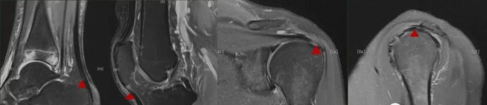
Degenerative changes? tear?
In this issue:
What is the Magic Horn Effect (MAE)?
Where does the magic horn effect occur?
How does the Magic Horn Effect perform?
Under what circumstances does the magic horn effect occur?
How to avoid the Magic Horn Effect?
What is the Magic Horn Effect?
Under normal circumstances, the direction of the arrangement of collagen fibers in each layer of tendon or articular cartilage is different, and it exhibits different properties in each direction, which is anisotropic; this tissue can change the movement of the water molecules in it, and the measured MRI signal will change with the change of measurement direction.
Water molecules and collagen fibers have a short T2 time under the action of dipole-dipole effect. Tendons exhibit low signal on MRI images; T2 time varies in the layers of articular cartilage, showing a layered performance.
However, in MRI imaging at a specific angle, these tissues can be manifested as a high signal of local abnormal elevation on a short TE imaging sequence, and the degree of increase of this signal is related to the angle of the main magnetic field B0, and when these structures and the angle of the main magnetic field are at 54.74 ° (about 55 °), the signal increase reaches the maximum, this phenomenon is called the "magic angle effect".

In the case of a fixed TE value, the MR signal strength changes with the change of angle within a certain angle width.
On short TE imaging sequences, it is characterized by an abnormally elevated high signal, the degree of increase of this signal is related to this angle, and the degree of signal elevation is maximized when the angle is 55°. This abnormally elevated signal is common on T1WI and PDWI sequences of short TE sequences, and normal tendons locally appear as high signals.
In MRI imaging, if the angle between the tendon and the main magnetic field is close to 55°, the locality of the tendon will appear as a high signal on T1WI and PDWI, but it will appear normal on long TE sequences such as T2WI. The presence of this magic horn effect may be confused with the pathological condition, causing a misinterpretation of MRI signal readings.
Where does the magic horn effect usually occur?
For the magic angle effect, you may be more familiar with the shoulder joint and Achilles tendon scanning; the magic angle effect often appears in the parts containing dense and anisotropic special tissue structures, such as tendons, ligaments and joint cartilage composed of collagen fibers, it should be pointed out that the magic angle effect is not limited to tendons and cartilage, it can appear in any part of the human body, such as MRI neuroimaging can also occur.
Under what circumstances does the magic horn effect occur?
Dense and anisotropic special tissue structures such as tendons, ligaments, and articular cartilage made up of collagen fibers.
The imaging structure is at a 55° angle to the main magnetic field.
USING SHORTER TE IMAGING SEQUENCES, SUCH AS THE T1WI AND PDWI OF SE SEQUENCES OR GRE SEQUENCES, IS MORE COMMON.
The horn effect can occur in any part of the tissue (e.g., shoulder, knee, ankle, wrist, hip, intervertebral disc, etc.) containing the above tissues (but not only in fibrin-containing tissues), in any orientation (transverse axial, sagittal, coronal).
How to avoid the Magic Horn Effect?
1. Increase the TE value appropriately. While te values aren't the only factor that determines the magic horn effect, changing it is the most effective factor in improving the magic angle effect.
Left: TE 35ms; the magic horn phenomenon is more obvious. Right: TE 70ms; no obvious magic horns.
In the direction of the magic angle, the T2 value of the tendon will be extended, and the result is an increase in MRI signal intensity when performing short TE imaging; this change in signal intensity is negligible when performing long TE sequence imaging.
At present, the scan of limb joints often uses sequence scans of short TEEs, such as T1WI, PDWI and intermediate weight sequences between PDWI and T2WI. Therefore, in order to avoid the misunderstanding of the MRI image signal caused by the magic angle effect, it is recommended to have at least one long TE sequence in the scanning sequence when performing joint scanning.
2. Change the scan position to avoid the magic horn effect.
When placing the scanning site, try to avoid the scanning site and the main magnetic field at an angle of 50 ° -60 °, such as the scanning of the knee joint and the Achilles tendon.
The magic angle effect is closely related to the angle of the main magnetic field, and multi-sequence multi-directional observation and comparison are required when interpreting the MRI signal in these parts, so as to effectively avoid the misunderstanding of the MRI signal. Most of the magic angle effect brings us artifacts that are not helpful to diagnosis; but at the same time, the magic angle effect can be used in some MRI special imaging of tendons, tendon sheaths, joint cartilage and other collagen-rich tissues, which can be conducive to the display and differential diagnosis of lesions.
bibliography:
Bydder G M . New Approaches to Magnetic Resonance Imaging of Intervertebral Discs, Tendons, Ligaments, and Menisci[J]. Spine, 2002, 27.S,
Regge,Zhang Guangfeng,Jia Huihui,Wu Jizhi,Chang Yan,Yi Peiwei,Sheng Mao,Yang Xiaodong. Research Progress on Short T2 Tissue Magnetic Resonance Imaging Technology[J].Chinese Journal of Medical Physics,2020,37(5):594-598.
Hashemi, Bradley, Lisanti, et al. MRI basis[M]. People's Medical Publishing House, 2019.
Source: MRI image
【Copyright Notice】This platform is a public welfare learning platform, reprinted for the purpose of transmitting more learning information, and has indicated the author and source, such as teachers who do not want to be disseminated can contact us to delete