1
Giant cell tumor of bone
Non-osteogenic connective tissue that originates from bones, the main component of the tumor resembles osteoclasts. Tumors are more common in young adulthood aged 15 to 30 years, mostly after the epiphysis has healed, but tend to occur distal to the femur, proximal tibia, and distal radius. In the early stages, there is only local intermittent pain. Often due to the development of tumors, local swelling, tenderness, or restriction of movement in adjacent joints. Larger tumors may have symptoms such as localized increased skin temperature, varicose veins, and tenderness.
Imaging presentation
1. Although giant cell tumors begin near the epiphyseal plate, they almost always grow over the epiphyseal plate and towards the joint, and mainly in this way, they can reach under the joint cartilage.
2. Most of the tumors are limited to the cancellous bone at the end of the bone and the epiphysis, and grow more to the original protruding part of the bone, so that the ward expands and expands, showing a lateral development. Its contours are often leafletized, and the cortex, although thinner, is still intact. There is no periosteal reaction and no tumor tissue mass around it. No tumor matrix calcification.
3. The tumor presents a clear density reduction area with normal bone demarcation, in which residual bone trabeculae similar to multi-atrial septum can be seen, and can be changed in a "soap bubble" when obvious.
4. In the early stage or advanced stage of the disease, its edges can be blurred, and if there is no trabecular bone in the middle, it is more difficult to diagnose, and it is generally based on the lesion site.
5. In the advanced stage of the lesion, excessive tumor swelling can rupture the bone cortex and can invade the nearby soft tissue.
MRI
T1 is mostly uniformly low signal, bleeding can show significantly high signal, T2 is mixed signal, low, medium or high signal is mixed, and tumor tissue is relatively high signal; significantly strengthened. The appearance of hemosiderin concentration and "bright spots" has certain significance for the diagnosis of bone giant.
Reading points:
1. It is more common in the young adulthood of 15 to 30 years old, mostly after the epiphyseal has healed
2. Adjacent joint surface growth, easy to develop to the bone process site
3. The long diameter is perpendicular to the backbone
4. The edges are not high-density hardened or more blurry
5. The bony interval is relatively thin
6. There is almost no periosteal reaction and calcification, and there is no osteogenesis

2
Aneurysmal bone cyst
It is more common in young people under 30 years of age; the site is more common with the long shaft epiphysis, pelvis, and spine; clinical treatment is mostly based on local swelling and pain; and there is a history of trauma. Pathologically, most of them are located in the middle of bone relaxants and medullar cavity, and a few are located in the cortex and periosteum; they are filled with dark red, brownish red and non-clotting blood; single or multi-room, with fibrooskeletal septum.
pathogenesis
1. Bone lysis stage: venous obstruction, increased intraosseous pressure, orthososis resorption.
2. Expansion period: higher intracapsular pressure, greater bone defects, piercing the bone cortex and lifting the periosteum.
3. Stabilization period: the intracapsular pressure is stable, and the blood cavity is separated by fibers and communicates with each other.
4. Healing period: calcium ossification in the capsule, thickening of the sac wall, and the formation of dense bone mass
Image representation
Eccentricity, blowout swelling, polyventricular or single-room osteolytic changes, thinning of cortex compression, clear boundaries with sclerosis. MRI sacs have a long T1 long T2 signal, fibrooskeletal septum T1T2 is a low signal, and there is a fluid level in the lesion (T1 is low and high, T2 is high and low), which is more characteristic.
Reading points:
1. It is more common at 10-20 years old, and it is more common for long bones to be dry-epiphyseal ends
2. The long diameter is parallel to the backbone
3. The joint surface is rarely affected
4. The swelling is generally more obvious, showing a "balloon-like blowing", the expansion can obviously see the periosteal reaction, and the sac interval can be calcified
5. Edge smoothness with hardening
6. There is a history of trauma
3
Chondroblastoma
Mostly located in the epiphyseal region (epiphyseal part of the epiphysis before the cartilage of the epiphysis closes); 1/3 of the cases occur in the tibia, the others are the upper and lower ends of the femur, the fibula, the distal end of the ulnar radius, etc.; 10-20 years old (60-80%), 5 years of age and above are rare.
pathology
Early stages: Chondrocytes are round or polygonal, the cells are tightly arranged, the nucleus is large, and giant cells can appear after necrosis.
Intermediate stage: increased calcification, degeneration and necrosis.
Late stage: the necrotic region is replaced by connective tissue, there are bone-like and even interosseous formations, and calcified ossification is significantly increased. Typically pane-like calcification.
image
Typically presents as an eccentric osteolytic destruction zone of the epiphyseum of a long bone, lobulated, well-defined, surrounded by a thin sclerotic ring with calcified spots.
MRI T1W shows a low signal, T2W is mostly mixed with high signal, cartilage-like matrix is a higher signal, calcifification is low signal, bleeding sac becomes high signal, and high signal is separated by low signal intervals. Pane-like calcification and high signaling of grape-like T2 are suggestive for chondroblastoma
Reading points:
1. 10-20 years old (60-80%), less common than 5 years old and over 60 years old
2. Limited to the epiphyseal or epiphyseal plate
3. There are many complete or incomplete thin layers of high-density hardening
4. No thick bone crest
5. Mild swelling, small lesions
4
Cartilage mucoid fibromas
Originates from the connective tissue of cartilage and consists mainly of mucoid cartilage. It is more commonly between the ages of 10 and 30 years, less common in people under 5 years of age, and less commonly in people over 60 years of age. It is more common in the lower extremities, especially in the upper tibial section, followed by the lower femur, the lower fibula, the calcane, humerus, and iliac. Clinical symptoms are characterized by intermittent pain, which ranges from several months to several years. A small number of patients are asymptomatic and are discovered incidentally after a physical examination or trauma. The tumor is pathologically round, oval, or lobular, with a smooth or convex surface. The tissue structure is mainly composed of three components: cartilage-like, mucus-like, and fibrous tissue, and the various components are somewhat variable.
Imaging presentation
The epiphyseal end of the long shaft (about 2 cm from the epiphyseal line) is eccentric cystic-like destruction, the long axis is consistent with the bone axis, and the edge is clear; there is often a hardening edge on the side of the medullary cavity, the wall of the capsule may have a bone crest deep into the capsule, the lateral bone cortex is swollen and thinned and wavy, and the calcification in the tumor is rare. Tumor signals on MRI vary due to differences in tumor composition, but mostly show abnormal signals of long T1 and long T2, and cartilage, mucus, and old blood are high signals, and there is significant strengthening after strengthening.
Reading points:
1. Most of the disease occurs between 10-30 years old, less than 5 years old, and less often over 60 years old
2. Long diaphyseal end of the backbone (about 2cm from the epiphyseal line)
3. The long axis is consistent with the bone long axis
4. There is often a hardened edge on the side of the medullary cavity, and the lateral bone cortex expands and thins in a wavy manner
5
Non-ossified fibromas
It is a benign tumor that occurs from the connective tissue of the bone marrow cavity, and there is no tendency to become osteogenesis, so it is also called "non-ossification". As bones mature, they may disappear on their own. Generally single-shot, occasionally multiple. The disease tends to occur in adolescents aged 8 to 20 years, with the highest incidence (29%) of the metaphyseal metaphyseal femur, followed by proximal tibia and both ends of the fibula. There are generally no symptoms in the early stages, and most are found after trauma. Rarely present with localized pain. Pathologically, the tumor is composed of tough, dense fibrous tissue and is encased in a bone shell. Microscopic tumors are composed of connective tissue cells, multinucleated giant cells, and foam cells. There is no osteogenesis in the lesion, and the surrounding bone tissue has reactive hyperplasia.
Imaging presentation
According to the X-ray performance, imaging can be divided into two types: cortical type and medullary type. The cortical type is characterized by lesions that are partial to one side of the diaphysis, located in or under the cortex, and are single- or multi-room, round, ovate, or lobular areas of light transmission with a coarse crest. The long axis of the lesion is mostly parallel to the backbone, and develops into the bone to protrude into the medullary cavity, surrounded by a dense sac shell or sclerotic band. Non-ossified. The medullary cavity type shows that the lesion is located in the center of the long bone, showing a single-sac or multi-cystic translucent area, the density is more uniform and there are sclerosis edges, the polycystic has irregular bony space or bone crest, and the local bone cortex is thinned.
MRI tumor components are collagen fibers, hemosiderin, etc., T2 is low signal, if it contains a large number of tissue cells, T2 is high signal; after strengthening, it is obvious. MRI tumor components are collagen fibers, hemosiderin, etc., T2 is low signal, if it contains a large number of tissue cells, T2 is high signal; after strengthening, it is obvious. If T1 and T2 are low-signal, they have certain characteristics and reflect the mature fibrous tissue inside the lesion.
Non-ossified fibromas are closely related to fibrous bone cortical defects, and most scholars believe that they are different stages of the same disease, that is, fibrous bone cortical defects before the age of 14, and after 14 years of age, without disappearing on their own, they develop into non-ossified fibromas, or ossified fibromas. Both have the same predisposing sites and similar histological manifestations. Therefore, we also believe that fibrous cortical defects often occur bilaterally symmetrically, generally disappearing spontaneously before the age of 14 years; if they do not disappear, they can develop into non-ossified fibromas. At the same time, fibrous cortical defects are more common in children aged 4 to 8 years, have familial tendencies, disappear spontaneously within 2 to 4 years, often appear symmetrically, appear in cystic or flaky bone defect areas, and have no swelling intrusion into the medullary cavity changes.
Reading points:
1. It tends to occur in adolescents aged 8 to 20 years
2. The epiphysis is partial to the backbone side
3. Mild dilated multi-room lesions
4. The diameter is greater than 2cm, and the long shaft is consistent with the backbone
5. No ossification, enlarged crest
6
Osteoblastoma
The dry epiphyseal end of the femur and tibia tends to occur in long bones; it is more common in children under 30 years of age; it is mostly a dull pain, and it cannot be relieved by taking salicylate drugs. The cause of the disease is that the blood supply is rich in the connective tissue matrix and its large number of osteoblasts and giant cells.
Image representation
The long bone is an oblong oval map-like bone destruction in the pulpolar cavity along the long axis of the backbone, which is marked by spots, cords or calcifications; the edges are clear and have sclerotic edges; eccentric growth accounts for 46%. MRI bone-like tissue T1 is low signal, T2 is high signal, calcifification or ossification in the lesion is low signal in each sequence, reactive hyperemia and edema can be seen around the area, enhanced bone-like tissue is strong, calcified, ossified, cystic, and bleeding areas are not strengthened.
Reading points: (cortical type)
1. 80% are under 30 years old
2. Good hair dry epiphysis, can develop to the backbone, bone end
3. The edge bone hardening is obvious
4. Light expansion
5. Spotted, cord-like calcification or ossification
7
Osteosa hemangioma
It can occur at any age, mostly in middle-aged people; the site is more common in the vertebrae and craniofacial bones, and long bones also occur. Pathological cavernous hemangiomas are mostly composed of large parenchymal blood vessels and blood sinuses, commonly found in the skull and spine; capillary types are composed of extremely dilated fine capillaries, most commonly found in the oblate bones and long shaft epiphyseal ends; there is often bleeding in the tumor to form blood clots or cystic cavities, and can also be organicized thrombosis and venous stones.
Image representation
Occurs in long bones mostly honeycomb or sac-like expansive bone destruction, soap bubbles, mesh, can be "dead branch" more characteristic. MRI is characteristic and presents as a streaked short T1 long T2 signal and liquefaction necrosis with a long T1 long T2 signal.
Reading points:
1. It is more common in adults, and the lesion is often located at the end of the bone
2. The lesions of long bones are soap-bubble-like and reticulated, and have certain characteristics of "dead branches"
3. There are more cortical swelling and hardening edges, and there are more bone ridges in the sac
4. MRI is characteristic and manifests as a striped short T1 long T2 signal
8
Ligamentoid fibromas
It is an extremely rare benign tumor. Growth is slow and well differentiated. The age of onset is mostly under 30 years of age, and there is no gender difference. The lower jaws are more common at the most common site, followed by the femur and pelvis. Ligament-like fibromas of long bones occur most often at the metaphyseal end. Clinical manifestations are predominantly pain and swelling. A few are found after pathological fractures. Pathologically, hard fibromas are gray, yellowish-white, hard, rubbery, and tough, and sometimes cystic changes can be seen inside the tumor. Microscopic tumors are composed of rich fibroblasts and a large number of collagen fibers.
Imaging presentation
Hard fibromas are mainly manifested by osteolytic osteolytic destruction, sclerotic rings are clearly visible at the edges, swollen and thinned bone cortex, and no periosteal reaction. Sometimes bone trabecular remaining in the bone destruction area and the soap bubble changes. Occurs in long bones mostly at the metaphyseal end, manifests as a central bone defect, and is distributed along the long axis of the bone, the bone cortex is thinned, and sclerotic bands can be seen around.
Reading points:
1. Mostly under 30 years old, mostly occurs in the epiphyseal end extending to the backbone
2. Visible hardening ring
3. Distribution along the long axis of the bone
4. The crest is large
5. MRIT1 and T2 are medium or low signals, the signal is more uniform, and the strengthening is not obvious
9
Brown tumors with hyperparathyroidism
Hyperparathyroidism called hyperparathyroidism, is due to the hyperparathyroid secretion of parathyroid hormone, causing calcium, phosphorus metabolism disorders of the disease, with a variety of clinical, but only some patients with bone changes biochemical tests can see increased serum calcium, serum phosphorus decreased, alkaline phosphatase plateau because of parathyroid adenoma or cancer, parathyroid hyperplasia bone changes include: osteoporosis, epiosteal cortex resorption, limited bone destruction and so on.
Imaging (brownoma)
Localized cystic bone destruction, manifested as sac-like translucent areas of varying size, individual, or multiple, with clear boundaries with surrounding bone, and the larger ones may swell outwards, sometimes with multi-room soap-like changes, similar to giant cell tumors, and are more common in long bones and mandibles. This single-room or multi-room cystic destruction is clinically called a brown tumor and can be repaired in a short period of time after surgical removal of the adenoma. Larger brownomas are often accompanied by pathological fractures.
Reading points:
1, easy to occur at any age, more common in women
2. Prone to pelvic, long bones, jaws
3. The long bone can be deformed, and the bone end is not widened, which can be accompanied by fracture
4. Biochemical examination can show an increase in serum calcium, a decrease in serum phosphorus, and an increase in alkaline phosphatase
10
Benign fibroushistiocytoma (BFH) is a mesenchymal tumor that originates in bone and belongs to the World Health Organization (WHO) classification of histiocytic-derived tumors. Fibrohis cell tumors are common in soft tissues, rarely originating in bone, but both have the same histological manifestations. Benign fibrohistoblastomas have fibroblast-to-histiocyte differentiation characteristics, similar to non-ossified fibromas at the metaphyseal end of the spine, but with different clinical and radiological features.
Clinical features: occurs in adults, and there is no significant difference in incidence between men and women. It can occur throughout the body, but most of the long bones of the limbs are used, especially the femur and tibia. Occurs more commonly in the epiphyseal ends. The remaining reports occur in the long bone backbone, pelvis, ribs, finger (toe) bone, cervical spine, and maxilla. Clinical manifestations are usually predominantly localized pain, sometimes with rational fractures. The disease is locally invasive, and can recur locally after surgical curettage, with progressive bone destruction but no metastasis.
Imaging features: lesions are osteolytic, with smooth borders, mostly sclerotic margins, and rough bone trabeculae visible inside. In long bones, it may originate from the diaphysis and the end of the epiphysis, which is more common in the epiphyseum. Initially, the lesion is located in the bone marrow cavity, eccentric growth, as the lesion progresses, the bone cortex is invaded and thinned, and pathological fractures can occur.
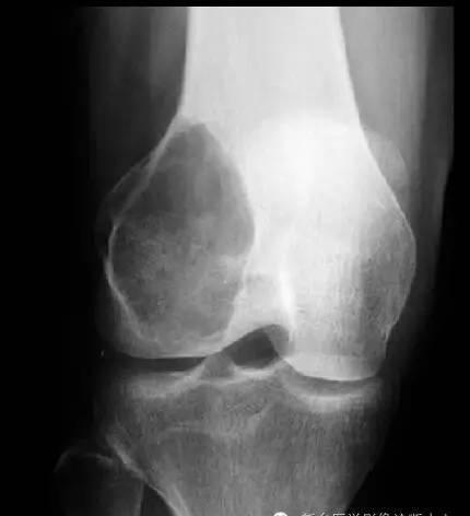
Clinical and imaging features: (for a picture analysis of bone benign fibrous histiocytoma, see figure above)
1. Age and medical history: female, 32 years old, right knee joint pain for one year
2. The site of the lesion: the right side of the femur epicondyle and the metaphyseal end
3. Number of lesions: single, eccentric
4. Type of bone destruction: map-like destruction, no obvious swelling, no bone crest in it
5. Edge of the lesion: the boundary is clear, and the sclerosing band is visible
6. Type of lesion matrix (component of tumor tissue): no bone and cartilage calcifications are seen in the mass.
7. Bone cortex changes: no damage is seen.
8. Periosteal reaction: no obvious periosteal reaction was seen
9. Soft tissue mass: not seen
This disease should be distinguished from the following two diseases:
Osteoglobus tumor: mostly expansive, eccentric bone destruction, multi-atrial bone destruction, thinning of the bone cortex, no osteosclerosis and periosteal reaction, occurs around the knee joint, involves the bone end, angiography mostly has mild to moderate increased blood transport, are different from benign fibrous histiocytoma; histology is mainly fibrous yellow tissue, swirling, visible multinucleated giant cells and foam cells, giant cells are larger and have more nuclei; However, BFH spindle cells and multinucleated giant cells are small in size, sparsely distributed, have fewer nuclei, and have more foam cells In this group of cases, they all occurred at the end of the bone, and the preoperative misdiagnosis of expansive osteolytic destruction with clear boundaries was bone giant cell tumor, and the diagnosis must be based on pathology.
Non-ossification fibroids: (1) Age of onset: Non-ossified fibromas are more common in adolescents under 10 to 20 years of age, and BFH is more common. (2) Site: non-ossified fibroma occurs in the epiphyseal end of the long backbone or the backbone cortex, most of which are located around the knee joint, eccentric, the bone cortex is slightly expanded and thinned, but the hardening ring near the side of the medullary cavity is uniform, not thickened, and there are more fine separations in the destruction area, and the degree of transmittance is higher; The incidence of bone BFH is mostly found in the long bone backbone, the metaphyseal end and the epiphyseum, pelvis, ribs, and also in the collarbone and cervical vertebral body [benign fibrohistoma tends to occur in the iliac and ribs (where non-ossified fibromas never occur), and can also be seen in the long backbone, mostly located in the center of the backbone, occasionally in the epiphysis]. (3) Clinical: non-ossified fibromas are often multiple, clinical symptoms are not obvious, no pain, pain after the occurrence of pathological fractures, there is a tendency to self-healing; BFH is painful and has a tendency to recur after surgical curettage. (4) Histology: The histological performance of the two is similar, but there are relatively more BFH foam cells. Epiphyseal fibrosis defect occurs in children, and adults can heal spontaneously, no clinical symptoms, occurs in the epiphyseal cortex, the lesion range is small, is a neoplastic lesion; if the lesion is large, involving the medullary cavity and forming a mass, it is a non-ossified fibroma.
Bone malignant fibrohistoma: mostly osteolytic osteopathic destruction, eccentric growth, unclear boundaries, no residual crest and sclerosis, cortical often interrupted defects, invasion of surrounding soft tissues; Cells have distinct polymorphic, atypical, and pathological nuclear divisions that are easy to identify. Benign fibrous histiocytoma of bone has a poor prognosis, is prone to recurrence after resection, and has very few atypical cases of lung metastases. Correct diagnosis of the disease is helpful for clinical treatment, expanding the scope of resection, and eradicating the disease.
Source: Xinxiang Medical Imaging Diagnostic Center
【Copyright Notice】This platform is a public welfare learning platform, reprinted for the purpose of transmitting more learning information, and has indicated the author and source, such as teachers who do not want to be disseminated can contact us to delete