Recently, the Sixth Affiliated Hospital of Sun Yat-sen University admitted a patient with a huge tumor of the breast, because of the secret illness and avoidance of medical treatment, delayed the condition, resulting in his tumor as large as a watermelon, ulceration and pus. The patient's illness is unbearable, the mental pressure is huge, and many hospitals are helpless. On the verge of despair, after repeated discussions by the multidisciplinary joint diagnosis of the hospital, it was unanimously agreed that the risk of removing the tumor was extremely high, but in order to let the patient survive, in the face of heavy pressure and problems, the team of director of breast surgery Li Haiyan never gave up, and finally successfully operated on the patient, so that the patient could regain a new life and return to a healthy life!
Initially the lump is only two centimeters and grows into a huge mass in 15 years
Ms. Xu, from Liwan, Guangzhou, is in her 40s and feels incredible when she talks about this huge tumor. As early as 15 years ago, the right breast lump was found, it was the third year after childbirth, the tumor was not large, only 2 centimeters, about the size of broad beans, and she did not have any uncomfortable feelings, so she did not take it seriously, and applied essential oils for maintenance.
Two years ago, she found that her breast lump began to increase rapidly, accompanied by local necrosis, ulceration, pus, odor, huge mass caused her to be unable to sit and walk, half a year ago began to be bedridden, the patient's spirit, sleep, appetite is very poor, emaciation is very obvious, the maximum diameter of preoperative mass has reached 50 cm, the quality of life has dropped sharply, bringing great burden and mental pressure to the whole family.
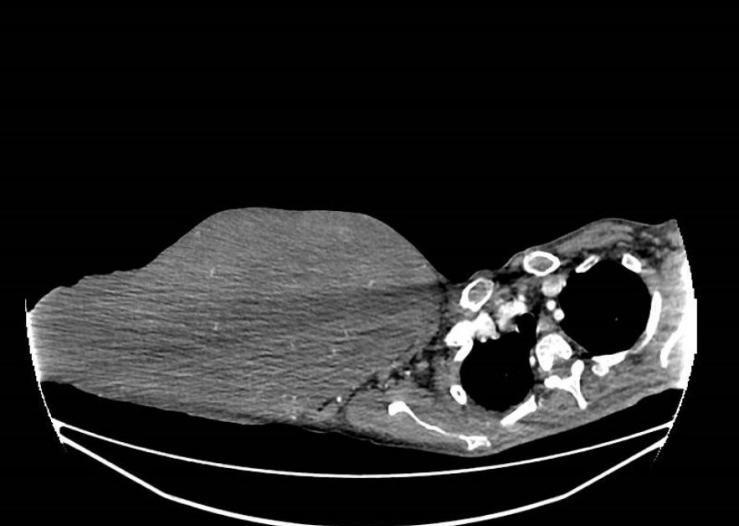
Chest enhancement CT image.
The maximum diameter of the right breast mass has been measured before surgery to 50 cm
In the past two years, the family has taken Ms. Xu to many hospitals, and they have been told that there is no chance of surgery. The family was disheartened, and after being recommended to find Li Haiyan, director of the Department of Breast Surgery of the Sixth Affiliated Hospital of Sun Yat-sen University. Further examination confirms the diagnosis of a huge mass in the right breast with ulceration, infection, severe anemia, and hypoproteinemia.
After admission, the ultrasound of the breast indicates that the right breast is huge and has a large tumor, which does not rule out the possibility of malignancy. CT of the chest suggests a large soft tissue density mass in the right breast, about 318x120x425 mm in size (upper and lower diameter). PET-CT testing shows no tumor metastases. Biopsy is performed for mass excision, and pathology suggests consideration of lobular tumors.
If Ms. Xu does not surgically remove it in time, she may compress the chest cavity and abdominal organs due to the further enlargement of the tumor, resulting in the failure of the compressed organs, and in more serious cases, the rupture of the tumor can cause heavy bleeding and endanger life.
Due to the poor general condition of the patient, after multidisciplinary consultation and discussion in the hospital, it was decided to perform a right breast macromastomy after daily wound change, anti-infection, correction of anemia, hypoproteinemia, and other symptomatic support.
Multidisciplinary teams join forces to cut through thorns and tear off huge "baggage"
In order to successfully and safely dismantle this giant "bomb", Li Haiyan, deputy chief physician of the breast surgery department of the hospital, Wan Yunle, chief physician of hepatobiliary surgery, liao Hongying, chief physician of thoracic surgery, and other multidisciplinary experts formed a "bomb disposal team", and formulated a thorough surgical plan and made full preoperative preparations before the operation.
Under the escort of the team of director Jin Sanqing of the Department of Anesthesiology, after disinfection, the patient's tumor rupture wound was wrapped in a sterile specimen bag, and the assistant fixed the tumor throughout the process, Li Haiyan, Wan Yunle and Liao Hongying took the incision surrounding the huge tumor at the surface 2 cm from the bottom of the tumor base, closely followed the surface flap of the free mass in the dermis layer of the skin, and encountered a number of thick and tortuous veins in the process of freeing, and carefully sutured to stop the bleeding. After nearly 3.5 hours of "stripping away the cocoon", he fought hard and finally successfully removed the huge tumor and lifted the crisis.
"After practicing medicine for so many years, it was the first time I saw such a huge tumor. The nutrition that the patients ingested was all supplied to this behemoth." When the tumor was removed, all the medical staff present were stunned, and even the chief surgeon Li Haiyan couldn't help but sigh.
The results of postoperative pathological examination showed that the right mammoma was 45x29x37 cm in size, and the microscopic histology showed that the tumor cell phiss-type diffuse infiltration, mild-moderate heterosexuality, considered as a benign tumor, and tended to benign lobular tumors are more likely. The huge breast mass was removed, and the patient's life was saved.

The huge lump cut weighed 24 pounds.
Doctors remind: if there is an abnormality in the breast, you should see a doctor in time
Breast tumors are the most common clinical manifestations of local painless masses, so female friends do not have a fluke mentality, every year should be regular physical examination screening, once found breast tumor clues, including skin red eczema, orange peel-like changes, breast lumps, nipple discharge, etc., should be timely to the regular hospital breast surgery department for treatment.
Breast lobular tumors originate from mesophyll tissue and are benign, borderline, and malignant, with a very low proportion of malignant. In Asian countries, the average age of patients with phylloxid tumors ranges from 25 to 30 years. The disease often presents clinically as a rapidly growing painless single tumor with an inconsistency of enlargement and malignancy, and rarely chest wall fixation. Li Haiyan said that for rapidly growing breast masses, it should be clearly diagnosed as soon as possible to avoid complications and increase the difficulty of treatment. The local recurrence rate of phylloid tumors is high. She reminded: The postoperative follow-up of leaf tumors is due to close follow-up. For 5 years after surgery, see the doctor every 3 months to 6 months for physical examination and imaging examination. After 5 years, see the doctor once a year.
Text/Guangzhou Daily, Xinhuacheng Reporter: Liang Chaoyi Correspondent: Jian Wenyang, Dai Xi'an
Photo / Guangzhou Daily, Xinhuacheng Reporter: Liang Chaoyi Correspondents: Jian Wenyang, Dai Xi'an
Guangzhou Daily Xinhuacheng Editor: Li Jin