Cardiovascular disease is the highest mortality rate in China, the first chronic disease, which seriously threatens the health level of the people. In the daily work of ultrasound, patients are often heard to say that they are uncomfortable to come to the doctor, at this time, it is particularly important to understand the heart color ultrasound. Cardiac color ultrasound, or echocardiography, is the examination and evaluation of the internal structure and function of the heart through ultrasound, and then guides early clinical intervention and early treatment. Today, I will talk about the problems related to cardiac ultrasound.
How does cardiac ultrasound work? Is there any damage to the human body?
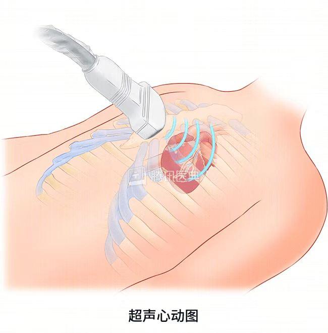
Cardiac ultrasound can use ultrasound scanning, through the human bones and muscle soft tissues to observe the heart structure, pulse and blood flow, etc., to clarify the function and integrity of the ventricles and large blood vessels, it is not radioactive and toxic, is a non-invasive examination technology.
What symptoms do I need to have a cardiac ultrasound?
When the doctor auscultates a heart murmur, considering congenital heart disease, valvular disease and other diseases, it is necessary to do cardiac ultrasound; people with coronary heart disease, especially those who have a history of myocardial infarction, must do cardiac ultrasound; when you have chest tightness, fatigue, less urine after activity, you must do cardiac ultrasound when you sleep and can't lie flat; when there is high blood pressure, hyperlipidemia and heart disease family history, the middle-aged and elderly also recommend regular physical examination, because many patients with atypical symptoms need to be examined for cardiac ultrasound to understand the extent of these diseases affecting the heart. Assistance with clinical treatment options.

Can a heart ultrasound confirm or rule out heart disease?
Cardiac ultrasound is mainly to observe the structure and function of the heart. For example, congenital heart disease and valvular disease can be diagnosed and excluded by heart ultrasound, and other heart diseases such as coronary heart disease, hypertension, arrhythmias, etc. Heart ultrasound can only provide information on the structure and function of the heart, and cannot rely solely on cardiac ultrasound to confirm or exclude heart disease, and other methods are needed to make comprehensive judgments. For some patients with congenital heart disease, it can determine what type of congenital heart disease it belongs to, so as to determine whether to operate and what the surgical plan is. For some patients with atrial septal defects and ventricular septal defects undergoing interventional occlusion, cardiac ultrasound can also be used to detect the appropriate location of the occlusion device during surgery.
What is the difference between cardiac ultrasound and ECG and coronary CT? Does cardiac ultrasound replace the other two?
Electrocardiogram, cardiac ultrasound, coronary angiography and coronary CT are all common methods used for the examination and diagnosis of heart disease. If the heart is regarded as a house, the cardiac ultrasound mainly looks at the structure of the house; the electrocardiogram mainly looks at whether there is a problem with the wires; the coronary CT mainly looks at the patency of the water pipe. In layman's terms, the cardiac ultrasound senses whether the hardware structure of the "house" is defective due to old age or other reasons through ultrasound, such as the size of the room, the effect of the partition wall, the stability of the doors and windows; the electrocardiogram can see whether the current of a house is regular and unimpeded, and whether there is a room current that is too chaotic and causes the bulb to burn out, which in turn affects the normal operation of the entire electric gate. Coronary CT and coronary angiography are examined for the patency and degree of obstruction of the aorta (coronary arteries), which can effectively judge whether there is narrowing of the coronary arteries; various cardiac examination methods have their own roles and advantages, and cannot be replaced by each other.
Since cardiac ultrasound is so important, shouldn't there be a heart ultrasound?
Cardiac ultrasound is not necessarily something that everyone has to do, and the following groups of people do not have to do cardiac ultrasound. When the previous cardiac ultrasound found that there is a slight reflux of the valve, when the auscultation hears a mild murmur, ultrasonography can not be done; there is no history of hypertension, heart disease, and now only mild chest pain, ecG can be preferred, and if necessary, heart ultrasound can be done; patients with coronary heart disease, no myocardial infarction, coronary CT examination is more meaningful than heart ultrasound; if there is a heart rate disorder such as palpitation and heart beating, the significance of doing ECG and 24-hour Holter electrocardiogram is greater than that of cardiac ultrasound.