If lung CT shows rapid deterioration and progression of lung lesions in the short term, tuberculosis and tumors are generally not considered. There is little literature on tuberculosis and tumors that are rapidly progressing in the lungs!
Today I share a case published in the journal BEST for nearly two years: 1 case of a fast-growing chest tumor in a young woman.
Figure 1 (Source: CHEST)
In the future, if anyone refutes you and says that the tumor will never progress rapidly, you can also use this case to face him with full confidence: it is possible!
Cases
38-year-old female, dry cough, progressive dyspnea, chest pain with nausea, vomiting, dizziness for 2 months.
There is a family history of ovarian, breast, and colon cancer. Family history will increase the incidence of tumors, patients in order to add fuel to the fire, once: a cigarette after a meal, race over the living immortals!
Physical examination: listlessness, shortness of breath, auxiliary muscle breathing, breathing 24 times / min, oxygen saturation of indoor air is 94%, left lung breath sounds disappear, and the rest of the body examination has no obvious abnormalities.
Laboratory tests
Lactate dehydrogenase (LDH) up to 1094 U/L;
Alpha-fetoprotein exaggerated to 33356 ng/ml (a common reference value in hospitals is.)
Human chorionic gonadotropin (β-HCG) is normal;
Blood calcium is as high as 17.6 mg/dl, which is the commonly used 4.39 mmol/L (calcium ion mg/dL × 0.2495 = mmol/L).
A chest CT 45 days ago is as follows:
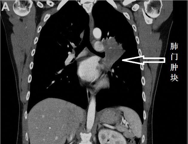
Figure 2 (Source: CHEST)
CT of the chest after admission is as follows:

Figure 3 (Source: CHEST)
In just 45 days, the previous lesion frantically occupied the entire left lung, and also invaded the pleura, causing a pleural effusion!
What strange disease?
Mediastinoscopic mediastinal lymph node biopsy, pathology: Nuclear protein in testis midline carcinoma, NMC, testicular nuclear protein midline carcinoma, referred to as midline carcinoma.
Figure 4 (Source: CHEST)
Shocked! Do women get the disease too?
Don't casually say how women without testicles can get "testicular nuclear protein midline cancer".
Many people do not know about this strange disease, because there are not many reports in the world, but the Guangdong Provincial People's Hospital reported 1 case.
Midline carcinoma of the testicular nuclear protein
Pathology is similar to squamous cell carcinoma in that it is a separate disease because of the rearrangement of testicular nucleoprotein genes, which is different from squamous cell carcinoma. In 2015, the World Health Organization (WHO) included it in a solid tumor of the chest.
The disease progresses very quickly. But there is almost no brain metastase, why?
It is speculated that the tumor is progressing with "lightning speed", and the patient has a short survival time and no chance of forming brain metastases.
If the tumor of lung cancer patients is widely metastasized throughout the body, but does not metastasize to the brain, it may be this strange disease.
So how is it treated? This patient lost the opportunity to operate in 45 days and did not give the surgeon a little "face" at all. Chemotherapy is not working well, and new drugs are still being studied.
What other fast-growing lung tumors are there?
Tumors generally do not grow rapidly, but in a few cases they do.
Scholars have reported a 28-year-old male, pulmonary inflammatory myofibroblastoma rapid growth, deterioration, huge lung mass occupies most of the right chest cavity and invades the superior vena cava, atrium and right pulmonary vein, adrenal glands, lymph node metastases. If the opportunity to operate is lost, inflammatory myofibroblastoma cannot be treated.
Rapid growth has also been reported for hamartomas.
Some scholars reported that 1 case of lung adenocarcinoma grew rapidly, and the lesion increased rapidly in only one month, and the authors considered that it was related to the giant lung vesicle.
Rapid growth has also been reported in lung sarcoma.
Scholars have also reported that patients with pneumoconiosis have rapidly growing masses, cut off as fibrotic lesions.
Some colleagues have encountered rapidly progressing diffuse lung adenocarcinoma, similar to heart failure, viral pneumonia, etc., and have not checked the literature and skipped it.
I previously met a patient, only 2 weeks, lung adenocarcinoma on the pleural metastases formed a pleural effusion.
epilogue
Midline carcinoma, accompanied by a rearrangement of testicular nuclear protein genes, progresses rapidly and as fast as lightning.
Tumors also occasionally mimic infectious diseases of the lungs.
bibliography:
[1] Yu Kuang Lai, MD; Brittany Holmes, MD; and Haiwei Henry Guo, MD, PhD. A Young Woman With a Rapidly Growing Thoracic Tumor. CHEST 2019; 155(5):e145-e148.
Chen Jingjing,Gao Xinglin. A case of primary pulmonary testonucleoprotein medium-line carcinoma and literature review[J]. International Respiratory Journal, 2018, 38(11):820.
[3] Suetsugu S , Yamamoto H , Izumi M , et al. [A case of rapidly growing inflammatory myofibroblastic tumor in the lung]. [J]. Nihon Kokyūki Gakkai zasshi = the journal of the Japanese Respiratory Society, 2009, 47(12):1156-60.
[4] Sagel S S , Ablow R C . Hamartoma: on occasion a rapidly growing tumor of the lung[J]. Radiology, 1968, 91(5):971-972.
[5] Nakamura H , Takamori S , Miwa K , et al. Rapid-growth Lung Cancer Associated with a Pulmonary Giant Bulla: A Case Report[J]. Kurume Medical Journal, 2003, 50(3-4):147.
[6] Sato S , Koike T , Yamato Y , et al. A case of rapidly growing pulmonary carcinosarcoma[J]. International Journal of Clinical Oncology, 2010, 15(3):319-324.
[7]大坂,喜彦, 渡邉,幹夫, 菊地,健. Rapidly growing progressive massive fibrosis that was difficult to differentiate from lung cancer: report of a case[J]. The Japanese journal of thoracic surgery, 2009, 62(6):504-7.
This article is reproduced from: Respiratory Channel of the Medical Profession
The author of this article is Sun Danxiong, a court of Yunnan Province
Image source: 123RF
Disclaimer: WuXi AppTec's content team focuses on the global biomedical health research process. This article is for informational purposes only and the views expressed herein do not represent the position of WuXi AppTec, nor do they represent WuXi AppTec's support for or opposition to the views expressed herein. This article is also not recommended for treatment options. For guidance on treatment options, please visit a regular hospital.
Share, like, watch, pass on new medical knowledge