Mr. Liu, male, 54 years old, found today that the head translucent nodules, mung bean size, no pain, no pain in pressing, did not put on the heart, but with the passage of time, the head and facial skin lesions became larger day by day, and the skin ulcer occurred recently, Mr. Liu gradually began to pay attention, came to the hospital for further help, after a series of physical examinations, combined with skin biopsy, and finally diagnosed as skin basal cell carcinoma.
If you have symptoms similar to Mr. Liu's, you need to pay attention to the fact that it may not be an ordinary skin disease, but cancer is coming to you.
<h1 class="pgc-h-arrow-right" data-track="40" > 3 abnormalities in the skin, alert to cancer</h1>
Through the analysis of Mr. Liu's case, it is not difficult to find that it has 3 more significant features:
(1) Translucent (pearl-like) nodules
Pearl-like nodules are typical of basal cell carcinoma lesions and are a key differentiator from other diseases, and if you find that this feature appears in the skin lesions at the exposed site, you should not be careless.
(2) There are signs of ulceration
Like our general skin diseases, such as freckles, melasma, sun spots, pigmented nevi, etc., are not easy to cause skin ulceration, if the skin appearance is small, you can not care, but if your skin lesions like the above cases, has begun to show signs of ulceration, then, please go to the hospital as soon as possible for examination, because cancer may be close to you.
(3) The lesion is located in the exposed area
Many factors play an important etiological role in the occurrence of basal cell carcinoma, of which ultraviolet exposure to sunlight is the most important environmental cause. In particular, people with fair skin, light eyes, red hair, elderly or childhood freckles, and a history of frequent personal sunburn have a higher risk of basal cell carcinoma associated with ultraviolet exposure, which is why basal cell carcinoma is more likely to occur in exposed parts of the skin. Like this Mr. Liu's skin lesions are on the face, where there is more opportunity to receive ultraviolet rays. Therefore, everyone should pay attention to sun protection in their daily lives and try to avoid long-term exposure of ultraviolet rays.

<h1 class="pgc-h-arrow-right" data-track="41" >What is basal cell carcinoma and what harm does it do to the human body? </h1>
Basal cell carcinoma is a common skin malignancy, belongs to non-melaninous skin cancer, more likely to occur in the elderly over 50 years old, there is no significant difference in the incidence of men and women, often occurs in the patient's skin is often exposed to the place, such as the head and face (especially the face is the most common, followed by the eyes, nose, etc.), at the beginning, there is no special performance, some people just appear skin bulge, most of them only the size of mung beans or even needles, it is easy to be ignored, but over time (less than a few months, More than a few years) the location of the skin lesion will have a scaly desquamation phenomenon, and then continuous scabs, desquamation, and then scabs, and then desquamation, skin ulceration, blood oozing on the surface of the skin where the lesion is located, and serious lesions will continue to expand, ulcers appear in the middle, and the edges are extremely uneven. Although the probability of metastasis is very small, its damage to local tissues is very serious, because it tends to occur on the head and face, and if it is not found and treated in time, it may even have the risk of disfigurement.
<h1 class="pgc-h-arrow-right" Data-track="42" What are the types of basal cell carcinomas >? What are the clinical manifestations? </h1>
(1) Nodule ulcer type: nodule ulcerative basal cell carcinoma is the most common type of basal cell carcinoma, more likely to occur on the face, its early clinical performance is mostly prominent skin wax-like nodules, small only the size of the needle, the larger one is about the size of mung beans, with the change of time, the skin lesions will increase day by day, pale gray or light yellowish brown hard nodules, at this time the skin lines on the surface of the lesion have basically disappeared, and the skin is thinner and thinner little by little until it ulcerates. Once the skin ulcerates, it becomes difficult to heal, and the bottom of the ulcer is granulated, cauliflower-like, granular growth, and the surface is also covered with a layer of secretions, which are serous. As the ulcers continue to enlarge, we can see many small nodules, which grow like pearls, uneven and ulcer-shaped around them. Some of the middle of the ulcer will heal into scars, and some will break, but the skin around it will continue to break to the edges, and at the same time invade the deep part, which can easily lead to disfigurement.
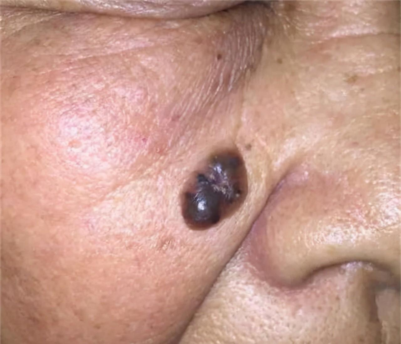
(2) Pigment type: This type of basal cell carcinoma, its appearance such as nodular basal cell carcinoma, however, as the name suggests, pigmented type, pigmentation will occur in the lesion, compared with the pale yellow and light gray of the nodular ulcer type, it appears to be darker brown or dark gray. This type is sometimes misdiagnosed as melanoma.
(3) Morpheular disease-like or fibrotic type: This type of basal cell carcinoma is relatively rare, accounting for only 2% of the total, and its age of onset is more youthful, more often occurring in young people, and can also be seen in children. Single hair is more, more likely to occur on the face. Its clinical manifestations are yellow-white waxy to sclerotic plaque, flat or mildly depressed, invasive growth, irregular shape and some are creeping, varying in size, some are only a few millimeters, some lesions are relatively large, and even the entire forehead can be occupied. Typical ulcers and crustings are absent and progress slowly, with smooth lesions in the early stages, and some small lesions are more easily overlooked until the late stages.
(4) Superficiality: This type is more common in men, overall is relatively rare, its age of onset is also biased towards young people, and most basal cell carcinoma is different, it is more often occurred in the non-exposed parts of the body, the trunk is more common, especially the back and chest, skin lesions have single hair, multiple, manifested as erythematous scaly patches, mild infiltration, its epidermis is thin, but the boundary is clear, the boundary is linear, slightly upward bulge, the middle of the lesion often has superficial ulcers and scabs, Some look like eczema, some look like psoriasis, need to be distinguished, its growth rate is slow, there are also pearl-like edges, this type of non-surgical cure often leaves a smooth atrophic scar.
In addition, there are other rare types, such as: mole basal cell carcinoma syndrome, fibroe epithelioma type, flat scar type, etc., because these types are relatively rare, their clinical manifestations are not described in detail.
<h1 class="pgc-h-arrow-right" Data-track="43" what are the diagnostic methods for basal cell carcinoma >? </h1>
Clinically encountered to the hospital for the diagnosis of suspicious lesions of patients to the hospital for treatment, generally the first step after seeing the patient is the collection of medical history, that is, we mainly have to figure out how the patient's skin lesions are found, how to find, how long it has been, and whether there have been any special changes recently to assist us in guiding our next step of examination, although this step is not the key to diagnosis, but it is indeed an indispensable link, and the cooperation of patients is needed at this time.
After an initial understanding of the patient's main condition, we usually need to conduct a physical examination of the patient. For patients suspected of basal cell carcinoma, we will focus on skin and lymph node examination, because the reason why the patient comes to the doctor may be only one lesion, and there may be other skin lesions in other parts of the patient's body that the patient has not noticed, which requires us to carefully examine to avoid missing a skin lesion due to poor examination, resulting in more serious consequences caused by untimely treatment.
At the same time, the examination results should also be recorded in detail, these are the factors and diagnostic basis for guiding the auxiliary examination, the main content that needs to be recorded is mainly the site where the skin lesion appears, and whether the size, color, shape and edge of each lesion are neat, how the texture, the degree of activity is, and whether there is an ulcer.
If patients with trans-lymph node metastases are found, the location, size, number, etc. of lymph nodes need to be recorded. Because basal cell carcinoma mostly occurs in areas where sunlight can be irradiated, it is necessary to examine the areas that can be irradiated by sunlight more carefully, and pay attention to the presence of precancerous lesions and other skin lesions in other areas.
Suspicious lesions found in the examination process should consider whether to carry out skin biopsy screening, some also need to be based on the situation of some other specific examinations, such as chest x-ray, B ultrasound, tumor markers, etc., but the pathological results are the gold standard for diagnosing diseases, only by clarifying the diagnosis of the disease, we can carry out the next step of treatment according to the diagnosis and personal disease characteristics.
<h1 class="pgc-h-arrow-right" data-track="31" > what are the treatments for basal cell carcinoma? </h1>
Since metastasis rarely occurs in basal cell carcinoma, there is a high possibility of cure with appropriate treatment of the primary lesion. Although there are several established treatments for primary lesions, their use varies depending on the risk of recurrence and the patient's condition. Therefore, pretreatment assessment of relapse risk factors is important in the choice of treatment. Clinical risk factors, including immunosuppression, previous radiation therapy for tumor lesions, tumor recurrence, poor border clarity, high-risk areas, etc.
Surgical resection is the standard treatment for basal cell carcinoma because it is very effective in achieving complete tumor resection at different anatomical sites. Microsurgery MMS on the Mohs allows for histological assessment of the surrounding edges during surgery. The tumor is removed layer by layer, and each layer is examined under a microscope until no abnormal cells remain. MMS is more effective at completely removing basal cell carcinoma than traditional surgical resection. However, this method is time-consuming and expensive than traditional surgical resection, which is more suitable for the treatment of tumors with a high risk of recurrence.
Electro-drying and curettage remove the surface of the tumor with a blade or scraper, and then burn the remaining base with an electric needle to control bleeding and destroy the remaining tumor cells. Therefore, it is more suitable for basal cell carcinoma in low-risk areas without invasive histopathological features.
Cryosurgery directly applied liquid nitrogen to the tumor leads to the formation of ice crystals outside and inside cells, thereby damaging the cellular phospholipid membrane and surrounding tumor-associated blood vessels, although simple, tumor resection is not precise and is not suitable for the treatment of tumors with a high risk of recurrence or the treatment of tumors that may lead to adverse cosmetic effects.
As an immune response modifier, imiquimod is used to induce immune cells to produce cytokines and stimulate cell-mediated immunity. However, miquimod is less effective than surgical resection. And imiquimod has more local side effects, so imiquimod is usually used for the treatment of superficial basal cell carcinoma at low risk.
The advantages of radiation therapy are aesthetic, however, basal cell carcinoma that recurs after radiation therapy may exhibit greater aggressiveness, including secondary recurrence and distant metastasis, as well as many side effects. In addition, good cosmetic results may deteriorate over time. Therefore, radiation therapy is usually only indicated for patients who are not suitable for surgical treatment, especially those whose tumors are located in high-risk areas.
In general, basal cell carcinoma has low metastasis potential and a slow clinical course. However, tumor infiltration may lead to recurrence. The risk of recurrence depends on a variety of factors, including tumor site and histopathological subtypes. Therefore, in order to select the optimal treatment strategy, it is necessary to conduct a pre-treatment clinicopathological assessment of the risk of recurrence. For treatment of the primary lesion, routine surgical resection is standard. However, depending on the tumor site, pathological subtype, patient condition, cosmetic effects, and treatment costs, other treatment options may also be appropriate.