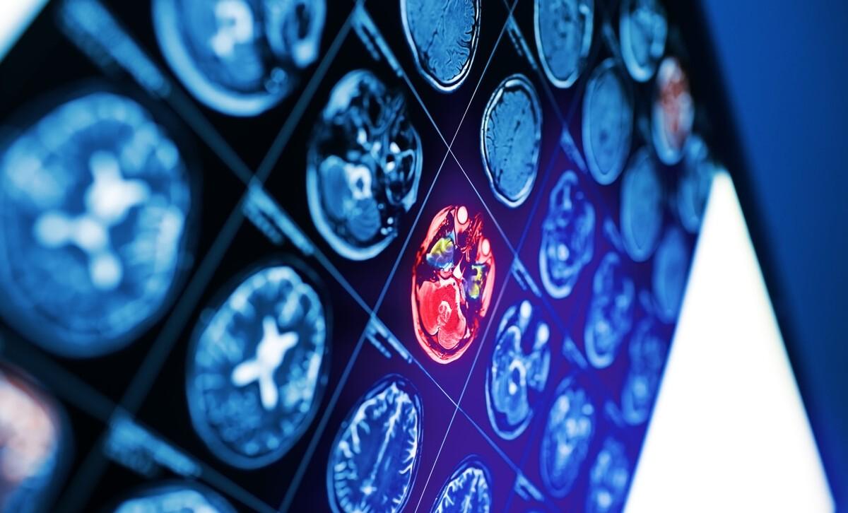
We have been in contact with magnetic resonance imaging (MRI) examination during the physical examination or medical treatment, and probably the most examined site is the head MRI due to headache and brain fever, and the brain MR examination report often sees "high signal of cerebral white matter", "lacunar cerebral infarction", "perivascular space enlargement", "multiple demyelination changes", "leukopenia" and even "cerebral ischemia lesions" such diagnostic reports, what is the clinical significance of these different diagnostic names?
Leukoaraiosis LA is a group of clinical syndromes with diffuse spots or patchy ischemic changes in the white matter of the brain around the ventricles and in the central semi-ovary region of the brain caused by a variety of different etiologies. With the widespread application of CT and MRI, LA is increasingly common in middle-aged and elderly people, but its exact cause and mechanism are not very clear, and its exact clinical significance needs to be explored. By CT or MRI scan, LA is described as diffuse leukopenia, a clinical syndrome described by a set of imaging agents caused by a variety of different etiologies. The main data on the pathogenesis of LA believe that ischemic injury is the most important factor in the occurrence of LA, followed by cerebrospinal fluid circulation disorders and permeability changes in the blood-brain barrier.
THE LESION OF LA IS MAINLY LOCATED IN THE WHITE MATTER OF THE BRAIN, WHICH IS ANATOMICALLY CHARACTERIZED BY IT ISCHEMIC. The lesion range of LA is mainly located in the cerebral white matter around the deep ventricles of the cerebral hemisphere, which is mainly supplied by the long medullary branch of the cortical and the deep perforating branch of the white matter, both of which are terminal arteries, coiled and spiral-like changes, with little or no collateral circulation, belonging to the watershed region, which determines that the white matter in this area is most susceptible to ischemia and causes chronic damage to blood vessels.
CT manifestations of LA: patchy or diffuse interventricular low-density foci on both sides of the cerebral cortex and per ventricles, blurred edges, moon halo-like, unreregulated, often symmetrical on both sides; often combined with bilateral ventricular enlargement and brain atrophy.

MR of LA: lesions showed low signal on T1WI, high signal on T2WI and T2flair, and the lesion site was consistent with CT, but more sensitive than CT, more clear to ventricular wall unevenness, and no reinforcement of enhanced scans.
LA is an imaging diagnostic term that is clinically symptomatic and can cause cognitive dysfunction, lower extremity dysfunction, and other cortical dysfunction. Imaging data suggest that the incidence of LA increases with age, and age and hypertension are currently independent risk factors for LA. Risk factors for the development of patients with cerebrovascular disease may be related to long-term hypertension leading to insufficient blood supply to deep white matter blood vessels, resulting in demyelinating of nerve cells and eventually manifesting as leukopenia.
According to the 2015 "Expert Consensus on the Diagnosis and Treatment of Cerebral Small Blood Vessel Disease" in Mainland China, the imaging of cerebral small blood vessel disease is mainly manifested as lacunar cerebral infarction, white matter lesions, vascular space enlargement and cerebral microbleeds. Therefore, LA is not in the category of cerebral small blood vessel disease, but it is closely related to the occurrence of small cerebral vascular disease. LA is indeed an important risk factor for the development of cerebral ischemic diseases, and the occurrence of LA in cerebrovascular patients is associated with small vascular lesions. Therefore, LA is both a consequence of cerebral small blood vessel disease and a risk factor for cerebrovascular disease, but LA cannot be regarded as one of the independent small blood vessel diseases.
It is generally believed that LA is a clinical syndrome described by imaging caused by a variety of different etiologies, and many causes of demyelinating, edema, ischemia, hypoxia, and microinfarction can cause abnormal changes in white matter, resulting in leukoplasmpor. LA is an imaging test result, and there are many reasons to pursue this result. LA is not only seen in patients with cerebral infarction, small cerebrovascular disease, hypertension, diabetes, neurodegenerative disease, traumatic brain injury, encephalitis, CO poisoning, diabetes, hyperaacymicuria, mitochondrial cell disease, methotrexate poisoning, leukodystrophy, etc., but also accounts for a certain proportion in the normal elderly. Therefore, CT and MRI only reflect the phenomenon of white matter imaging and do not necessarily have pathological significance. It is only an imaging change corresponding to various pathological conditions. However, "ischemic lesion" is not a formal diagnostic term, in textbooks and medical literature search engines can not retrieve its definition and the corresponding theoretical basis, it can indicate a state of ischemia and hypoxia, and defined as "ischemic foci" can only be regarded as an imaginary oral description or common name. Clarifying the cause of the imaging results of high white matter signal in the brain will have different plans and measures for preventing the occurrence of LA. In the past, people always attributed LA to cerebrovascular disease, and through this discussion, I hope to have a new understanding of LA