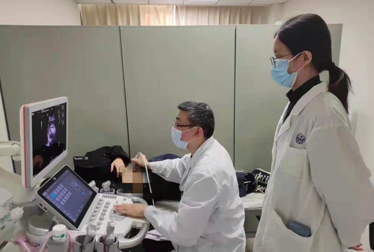
Director Jiang Lixin gave the patient an ultrasound examination
On December 2, when the ultrasound diagnosis report was obtained, a large stone in the heart of the 42-year-old patient, Ms. Zhang (pseudonym), finally landed.
"For nearly a year, I have been worried that the lump in my spleen is a malignant tumor, and I am so anxious that I can't sleep steadily. Now that I have finally been diagnosed with a benign problem, my heart is fixed! I would like to thank Jiang Lixin, director of the Department of Ultrasound Medicine of Renji Hospital, for their efforts for me. Ms. Zhang said.
Ms. Zhang has always paid attention to her physical health and will undergo a physical examination every year. During a routine physical examination last year, ultrasound examinations revealed low-echo clumps of about 36 mm in diameter in the spleen. Further MRI studies revealed that nodular abnormalities of about 3.6cm in diameter were visible in her spleen, and that they were significantly strengthened with a significant and progressive strengthening, taking into account splenic hemangiomas. Since the patient usually has no obvious clinical symptoms, the receiving physician at the time asked the patient to follow up closely.
There has always been a big lump in the spleen, which has almost become Ms. Zhang's heart disease. This year's physical examination, Ms. Zhang specially requested CT examination. The results suggested that the spleen occupied a significantly larger position than last year. Review of B timeout also found that the low echo clumps in the spleen were significantly larger than last year, with a diameter of about 48 mm, raised outwards, and seemed to be under-defined with the diaphragm, and malignancy was considered.
Could this lump be a malignant tumor? Ms. Zhang was extremely anxious, almost to the point of not being able to sleep at night.
On December 2, she went to Jiang Lixin, director of the Ultrasound Medicine Department of Renji Hospital, hoping to clarify the nature of the lump. While comforting her not to be too anxious, Jiang Lixin performed an ultrasound examination and found that the spleen was a low echo clumps, about 48 * 37 mm in size, and slightly raised outward.
"Although the mass is closely related to the diaphragm, carefully moving the probe can find that the mass and the diaphragm are relatively moving, and the demarcation is relatively clear." Color Doppler ultrasound shows that there is no obvious blood flow signal in the upper part of the mass, while the lower part of the mass is rich in blood flow signal, seeing a coarse blood vessel entering the mass from the periphery of the mass, coupled with the fact that the spleen has been occupied for more than a year in this case but there is no discomfort symptoms..." Jiang Lixin believes that considering benign or borderline tumors is more likely, and the possibility of vascular origin is large.
To further clarify the source of the mass tissue, Jiang Lixin performed an ultrasound for the patient. The results showed that the mass was rapidly enhanced compared with the normal spleen tissue, excluding hemangioma and hamartoma, and the mass was uniformly and highly enhanced after reaching the peak, which did not meet the characteristics of obvious bleeding and necrosis in hemangiosarcoma, so the possibility of hemangiosarcoma was ruled out.
In order to relieve the patient's distress and "verify the right body" of this mass, Jiang Lixin finally found a suspicious point by repeatedly playing the images under ultrasound: the mass was faintly visible in the arterial phase of a small, narrow strip of low-enhancement band, showing a radial change from the middle to the surroundings, a closer look, it turned out that the mass was relatively close to the diaphragm, with the patient's respiratory thoracic rise and fall, the imaging of the spleen caused interference with the movement artifacts, making these extremely small, Narrow strips of low-boost bands are not clearly shown by respiratory disturbances.
The central scar in the center of the mass, combined with radial narrow strips, forms a "spoke sign" structure. Based on this finding, Jiang Lixin believes that the mass belongs to a benign nodular lesion of the vascular tissue within the spleen and can be diagnosed as "sclerosing hemangioma-like nodular transformation (SANT)".
Jiang Lixin introduced that SANT is a very rare non-tumor, benign, angioproliferative lesion of the spleen, and there are currently few imaging data of the disease, and the literature at home and abroad only appears as a case report. The disease tends to occur in middle-aged women, most of whom are asymptomatic, with occasional cases of abdominal pain. The "spoke sign" in ultrasound images is a specific manifestation of the disease and is of great value for a definitive diagnosis. Although SANT is a rare benign lesion, a considerable number of patients have their blood systems affected, such as anemia and wasting, and surgery is the only radical cure for the disease.
On 4 December, the patient underwent surgery and the pathological diagnosis confirmed the correctness of the previous ultrasound diagnosis. The patient quickly recovered and was discharged from the hospital and embraced a healthy life again.
Author: Tang Wenjia
Editor: Tang Wenjia
Editor-in-Charge: Jiang Peng
Source: Hospital
*Wenhui exclusive manuscript, please indicate the source when reprinting.
![When the spleen is weak, the body will have 4 symptoms, which need to be observed carefully, and after discovery, timely conditioning[fig]](data:image/gif;base64,R0lGODlhAQABAIAAAP///wAAACwAAAAAAQABAAACAkQBADs=)