Conquer bone tumor imaging, maybe just a word
1. Bone cyst:
Proximal to the humerus or femur, central lesion near the growth plate with sclerosis margin, most likely a simple bone cyst. Low-density lesions on the side of the talus are most likely simple bone cysts. Typical signs: bone fragment fall

2. Chondrosarcoma (conventional type):
Adults (>30 years) + pelvis and long bones + expanding destructive lesions in the medullary cavity + popcorn in the tumor matrix / ring / comma calcification + cortical hyperplasia, intimal scallop-like changes + soft tissue masses may be present
3. Cartilage mucinoid fibroma:
10-30 years old + lower limb bone + adjacent growth plate + multi-chamber honeycomb + eccentric translucent lesion + scallop-like sclerosis edge + bone stack formed by new periosteal bone + flat tablet inconspicuous calcification foci + MR signal mixed
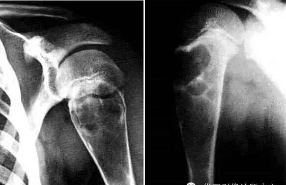
4. Benign osteoblastoma:
11-30Y + spine (spinous process, vertebral arch and transverse process) and long duct bone (metaphyseal end) + bone localized expansive low-density area + thinning or even rupture of the bone cortex + clear shell calcification around + spotted or large calcification or ossification within the tumor + generally no periosteal response.
5. Chondroblastoma:
Adolescents 10-20Y, limb long tube bone epiphyseal region, humerus, upper femur and proximal tibia are prone to, long bone epiphyseal end round, oval bone destruction, edges of different degrees of hardening, its common spots, flaky calcification
6. Non-ossification fibroma:
Proximal cortical lesions (site) of the distal posterior femur, with fan-shaped and sclerotic edges (morphology). The lesion is located on the medial side of the epiphyseal diaphysis of the long diaphysis, 3 to 4 cm from the epiphyseal plate, and 1 to 5 cm in size. Round or oval, eccentric growth, consistent with the bone axis, is lobed, thinning of the bone cortex, and the edge of the medulla is hardened and clearly demarcated. The bone at the border between the base of the tumor and the medullary cavity is slightly hardened, often soap foaming, and may be combined with pathological fractures.
7. Endogenous chondroma:
Popcorn-patterned, ring-shaped, punctate calcification foci + lobular growth + scallop-like edges of the endometrial surface of the concomitant bone cortex; short bones may be free of calcification
8. Giant cell tumor of bone:
20-40Y, after the growth plate is closed, occurs in the osteolytic lesions of the bone joint end of the bone without sclerosis, + saponaria sign, + eccentricity = most likely giant cell tumor; the cause of intracellular separation of bone giant cell tumor = bone wall crest + residual bone + connective tissue septal calcification
9. Osteoid osteoma:
Worsening pain at night + salicylic acid (aspirin) relief + LMD nest (center sclerosing) + peripheral reactive osteosclerosis
10. Spinal myeloma:
Multivertebral lesions + diffuse osteopathic destruction + osteoporosis + early nearby involvement, late involvement + vertebral body generally no kyphosis + less soft tissue masses + laboratory tests
11. Spinal metastases:
Primary history + multivertebral lesions + invasive changes + adnexal involvement + soft tissue mass + kyphosis + adjacent intervertebral space and intervertebral discs are generally undamaged.
12. Osteosarcoma:
A little breakthrough, full blooming: that is, osteosarcoma breaks through the cortex and forms a significant soft tissue mass.
15-25Y (75%), long shaft epiphysis (femoral, tibia); over 50 years old, more likely to occur in flat bones (iliac bones), ivory, cotton wool, needle tumor bone formation + insect phagocytosis, sieve-like bone destruction + lamellar, needle or radial periosteal reaction + periosteal triangle (Codman triangle) + soft tissue mass + tumor bone or neochondral calcification + osteolytic lesions show a slightly low to moderate signal on T1WI and moderate to high signal on T2WI. Osteoblastic lesions show low signal on both T1 and T2, and periotumoral edema is high signal on T2
13. Juve's sarcoma:
Adolescents (10-20 long bones; 20-25 flat bones) + invasive osteoclast + butterfly cortex + invasive periosteal response (periosteal triangle and radiation bone needles) + soft tissue masses
Source: Huaxia Imaging Diagnostic Center
【Copyright Notice】This platform is a public welfare learning platform, reprinted for the purpose of transmitting more learning information, and has indicated the author and source, such as teachers who do not want to be disseminated can contact us to delete