"Go to the hospital and take an MRI."
Have a small partner hear this sentence on the heart of a "giggle", always feel that this "nuclear magnetism" has a great harm to the body.
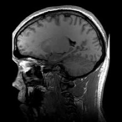
In fact, this "nuclear magnetic" that is, magnetic resonance imaging (MRI) is the use of nuclear magnetic resonance (NMR) principle, according to the release of energy in different structural environments within the material at different attenuation, through the additional gradient magnetic field detection of the emitted electromagnetic waves, you can know the position and type of atomic nuclei constituting this object, according to which you can draw a structural image of the interior of the object.

Magnetic resonance imaging of the longitudinal slice of the human brain, source Wikipedia
The "nucleus" of MRI actually refers to the hydrogen nucleus, and about 70% of the human body is composed of water, and the technology relies on hydrogen atoms in water.
Considering the patient's fear and fear of the "nuclear", doctors often refer to this technique as "magnetic resonance imaging".
Magnetic resonance imaging is often used in medicine to detect and diagnose heart diseases, cerebrovascular accidents and vascular diseases, detection and diagnosis of organ diseases in the chest and abdominal cavity, diagnosis and evaluation, tracking of tumor conditions and functional disorders, and diagnosis of exercise-related injuries.
In addition, magnetic resonance imaging (MRI) is often used in the detection and diagnosis of diseases of the reproductive system, breast, pelvis, and bladder because there is no risk of radiation exposure.
Compared with ordinary X-rays or computed tomography (CT), magnetic resonance imaging is one of the few clinical diagnostic methods that do not harm the human body and is safe, fast and accurate.
The "great hero" of this technology is the American chemist Paul C. Lauterbur, who won the 2003 Nobel Prize in Physiology or Medicine.
Paul C. Lauterbur, Credit: Ganga Library
Born in 1929 in the small town of Sydney, Ohio, Paul Lauterber received his B.S. from Case Polytechnic Institute in 1951 and his Ph.D. in chemistry from the University of Pittsburgh in Philadelphia in 1962. From 1963 to 1984, Paul Lauterberg was a professor in the Department of Chemistry and Radiology at the State University of New York at Stony Brook. He died on March 27, 2007 in Urbana, Illinois, U.S.A. at the age of 77.
Lauterbur developed a great interest in chemistry from an early age, and as a teenager, his parents helped him build a laboratory of his own in the basement.
When he was in high school, his teachers granted him permission to complete the experiments they wanted to do in class with some like-minded classmates, and granted exemptions if the experiments went wrong.
At a young age, he had "experimental freedom".
It is these young experiences that have allowed him to maintain a strong interest and unremitting efforts in the future on the road of chemical research.
Napkin graffiti
Such a high-end research on magnetic resonance imaging was originally born on a small napkin.
When Lauterberg was a researcher at the Mellon Institute of Industry, he dined at a restaurant on the outskirts of Pittsburgh, during which he brainstormed and scribbled the first model of MRI on napkins.
NMR (Nuclear Magnetic Resonance) is the scientific principle behind MRI, whose discoverers, Felix Bloch and Edward Purcell, were awarded the Nobel Prize in Physics in 1952.
However, in the decades that followed, magnetic resonance was mainly used to study the chemical structure of substances. It wasn't until the 1970s that Lauterberg and Peter Mansfield developed this research so that MRI could be used to generate images of the body.
Late-night experimentation
During his tenure as a professor in the Department of Chemistry and Radiology at the State University of New York at Stony Brook, Lauterberg devoted himself to the study of nuclear magnetic resonance spectroscopy and its applications.
Before him, most scientists placed specimens in uniform magnetic fields where radio signals were excitated from the sample as a whole. Lauterberg realized that if an uneven magnetic field was used, it was possible for radio signals to be excited from different regions of the specimen, potentially producing two-dimensional images.
At the time, the NMR at the State University of New York was shared by chemistry professors, and other professors made their measurements in a uniform magnetic field. To do this, Lauterbur had to do the research in the evening and restore the machine to its original setting the next morning.
Such a "night battle to pick the lights" laid the foundation for Lauterbur's research.
Submit with "clams"
After the success of the experiment, Lauterbur took the first photographs, including a 4 mm diameter clam, green pepper and two heavy water test tubes in a regular beaker collected by his daughter on the beach of Long Island Bay (the last one is particularly important because the human body is mainly made up of water).
Lauterbur then published his paper and his findings for the first time in the journal Nature, but the images in the paper were so vague that the journal editor rejected his request. But Lauterbur did not give up and continued his research. After repeated requests to the publisher for a second review, the paper was published. Later, the journal Nature rated the paper as one of the classic papers.
2/3 of the body weight is water, and such a high proportion is the basis for magnetic resonance imaging technology to be widely used in medical diagnosis. The water in various organs and tissues in the human body is different, and the pathological process of many diseases can lead to changes in the form of water, which can be reflected by magnetic resonance images.
On 6 October 2003, the Karolinska Institutet in Sweden announced that Paul Lauterber and Peter Mansfeld had been awarded the 2003 Nobel Prize in Physiology or Medicine in recognition of their groundbreaking achievements in the field of diagnosis and research in medicine.
The popularity of this technology has saved many patients' lives.
Modern Clinical High Field (3.0T) MRI Scanner, source Wikipedia
The emergence of new medical technologies is undoubtedly a blessing for all mankind, and the scientists behind the technology are the creators of the gospel.
The universe of science is vast, and the spirit of exploration of scientists is like the stars in the universe. The obsession and dedication to scientific research deserves our memory and praise.
END
Editor-in-charge/Heart & Paper
Swipe left to view the new media communication system of the Beijing Association for Science and Technology