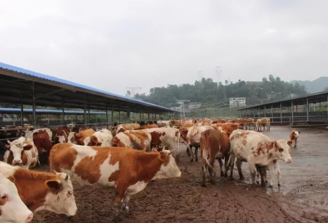
Bovine coccidiosis is one of the common parasitic diseases in the cattle industry, and cattle are susceptible to infection at all stages, but calves are more susceptible to infection. Because the disease is contagious, sick cattle should be isolated and treated as soon as they are discovered. At the same time, disinfect the environment exposed to cattle, strengthen management, and avoid the spread of disease.

First, understand the pathogen
There are more than ten species of Bovine coccidiosis: Coccidium Aymilia Chia, Coccidioides Amy, Leclavidae, Omelia Ahmed, Elliptera, Columnar Coccidioides, Canadian Coccidiosis, Oberamer Coccidioides, Araba Amy Coccidioides, Subspheres Amy Coccidioides, Brazilian Emmy Coccidioides, Eddie Amy Coccidioides, Omin Amy Coccidioides, Pilita Amy Coccidis, etc. Among the various coccidiosis that parasitize cattle, several of them, Amy Coccidioides S. coccidioides are more pathogenic and common.
Amy's coccidioides live in the rectal epithelial cells of cattle and can sometimes parasitize the cecum and lower colon; The oocyst is round or slightly oval, the egg wall is smooth, and the average size is 14.9--20 microns. Lecoccus spp., parasitic in the intestines of cattle; The oocysts are ovoid in shape, with an average size of 19.6 to 34.1 microns. Coccidiosis does not require an intermediate host for development. When cattle have swallowed infectious oocysts, the spores escape from the intestine into the epithelial cells of the parasitic site for split reproduction, producing fission in; Fission forms large and small gametes by gamete reproduction when it reaches a certain stage of development,
Large and small gametes combine to form oocysts excreted from the body; Oocysts excreted outside the body Spore in reproduction under suitable conditions, forming spore-shaped oocysts, spore-shaped oocysts are infectious.
Second, the identification of bovis disease
Diagnosis of bovis disease requires saturated saline flotation, microscopy of supernatant fluid, or direct microscopic examination of rectal scrapes, and the diagnosis can be confirmed by coccidioid oocysts.
Bovis coccidiosis is distinguished from other enteritis diseases, among which the easily confused one is E. coli infection, and after infection with E. coli, bovine diarrhea, feces with blood and odor, but more often in 10-day-old calves. After Salmonella infection, cattle are emaciated, the mortality rate reaches 50%, and as the disease progresses, joint enlargement occurs, sometimes manifesting as bronchitis and pneumonia.
Third, timely treatment of sick cattle
In order to avoid infection with bovine coccidiosis, it is necessary to take comprehensive methods such as isolation, treatment and disinfection, because adult cattle often have coccidiosis, so they should be raised separately, and the sick cattle are immediately isolated.
Bovine coccidiosis infection should be treated promptly for symptomatic treatment of sick cattle.
In the treatment of this disease, monensin, ampheraline, sulfamethazine are commonly used, and an appropriate amount is taken for internal treatment of sick cattle.
Fourth, strengthen management
1. Raise in groups to avoid contamination of feed by coccidioid oocysts;
2. The feces and mat grass of the barn-fed cattle need to be disinfected centrally or fermented by biothermal compost, and 1% keliaolin can be used to disinfect the barn and the feeding trough once a week at the time of illness;
3. Cow udders contaminated with feces should be cleaned before breastfeeding;
4. Add drugs to prevent bovine coccidiosis, such as amphetamine, according to the concentration of O.004% to 0.008% added to feed or drinking water.