Identification of microbial taxa of murals excavated from Nanliwang Village, Chang'an, Shaanxi
Cultural Relics Protection and Archaeological Science, No. 01, 1997, Guo Ailian, Yang Wenzong
Abstract The microbes excavated from the murals in Nanliwang Village, Chang'an, Shaanxi Province, were preliminarily analyzed and identified, and the results showed that the number of bacteria and molds was half that of each, and there was one kind of actinomycete. The main microbial groups are 8 molds, and 7 bacteria groups, a kind of streptomyces.
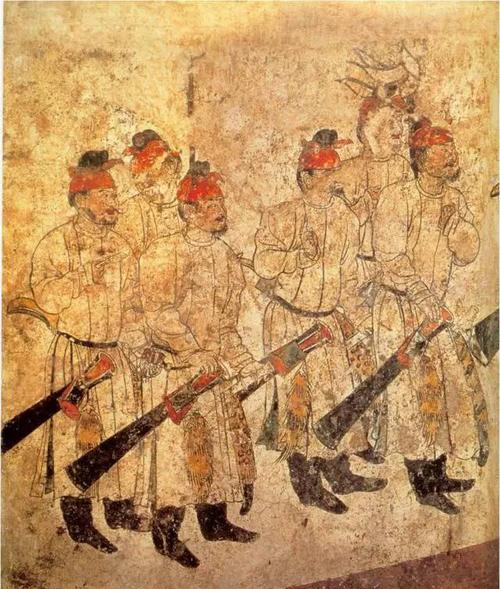
In the early 1980s, Shaanxi cultural relics archaeologists excavated the Tang Dynasty Wei family cemetery in Nanliwang Village, Chang'an County, adjacent to Xi'an South, and unearthed a number of Tang tomb murals. A few years later, we protected the murals. When it was repaired, it was found that the picture was covered with black and brown spots. Microorganisms are an important cause of this phenomenon, they blur the picture, and its harm is quite serious.

According to the previous analysis of mural pigments and materials, and the study of mural production processes, murals are not properly preserved, and there will be microbial contamination.
The production of Tang Dynasty tomb murals is carried out in strict accordance with certain processes, including wall treatment, drafting, finalization, and coloring. Generally after the completion of the tomb, the creation of murals began, and the walls were divided into earthen walls and brick walls. When painting murals on earthen walls, the walls need to be bulldozed first. When painting murals on brick walls, mud is used to clog and smooth the brick seams. Then smear the wheatgrass mud on the earthen wall or brick wall as the base, and when the wheatgrass mud dries, the picture begins. The picture is soaked in water with the white ash after the sieve plus the cut short hemp fiber, and fully stirred to form a paste, smeared on the base of the wheatgrass mud, the Tang Dynasty mural pigments are mainly natural minerals, some are simply processed by minerals, and a small amount of plant pigments are also found in some pigments. Generally, these pigments are mostly powdery, can not be directly painted, must be made of leather glue, fish glue and water.
Considering the mural making process, the presence of various glues in wheat grass, hemp fibers and pigments has created conditions for microbial survival. After excavation, it was exposed to the air, and the tomb was dark and damp, sometimes reaching more than 80%. So even inside the tomb microbes will spread.
Tang tomb murals are difficult to preserve in the tomb, and there is a possibility of collapse at any time. In order to protect the murals, it is currently better to remove the picture and place it in a museum with good preservation conditions. When uncovering the mural, first use a white cloth to paste on the picture to be revealed to stabilize the picture from falling off. The adhesive used is a natural resin, peach gum. The unveiled fresco is clamped with two plates covered with cotton or foam. Because it is difficult to repair after retrieval, microorganisms will grow over time.
Metabolites such as enzymes and organic acids produced by microorganisms during the growth and reproduction process are secreted from the fungus body to the material, causing the lime layer at the bottom of the mural to peel off, and the color also falls off. The pigment produced by microorganisms can cause color changes, so it is very important to stop the production of microorganisms in the mural and clean up the spots on the fresco pictures that have already affected it. In this paper, the microorganisms produced on the murals are analyzed and tested, their classification status is understood, effective technologies for preventing microbial contamination are established, and appropriate prevention and control methods are adopted.
1 Materials and methods
1.1 Sample collection samples were taken from the patches of mural pictures excavated from Nanliwang Village, Chang'an County, Shaanxi Province.
1.2 All used in the medium experiment is sterile normal saline.
1.2.1 Beef paste protein hypothesis medium (for isolating and culturing bacteria) Beef paste 3g, protein dolphin 10g, NaC15g, agar 20g, water 1000ml, pH 7.2, 0.IMPa sterilization 25min.
1.2.2 Tscher's medium (1) (for isolation and culture of mold) NaNO32g, K2HPO4 Ig^KCl 0.5g, MgSO4 0.5g, Fe-SO4O.Olg, sucrose 30g, agar 20g, water 1000ml, pH natural, 0.05MPa sterilization 20~25mino
1.2.3 Gao's synthesis No. 1 medium (for isolating and culturing actinomycetes) soluble starch 20.0g, KN031.0g, K2HPO4 0.5g, MgSO4-7H2O 0.5g, NaCl0.5g.FeSO4-7H2O (10%) has been dripped, agar 20g, distilled water 1000ml; pH 7.2~7.4, 0.IMPa sterilization 25mino
1.2.4 Nitrogen-free medium (for identification) mannitol 10g, KH2PO40.2g, MgSO4*7H2O 1.2g, NaCl 0.2g, Ca-SO4'2H2O 0.2g, CaCC) 35.0g, agar 20g, steamed water 1000ml, pH 7.2, 0.05MPa sterilization 20mino
1.2.5 Glucose oxidative fermentation medium (for identification) protein 2g, NaCl5g, K2HPO4 0.2g, W syrup 1%, washed agar 5~6g, l% aqueous solution of thymol blue 3ml, steamed water 1000ml, pH 7.0, 0.05MPa sterilization for 25min.
1.2.6 Glucose fermentation medium (for identification) beef paste 3g, protein dolphin 10g, NaCl5g, glucose 10g, water 1000ml, pH 7.2, add 20ml of cresol violet 0.04% aqueous solution, 0.05MPa sterilization for 25min.
1.2.7pH4.5 growth medium (for identification) with beef paste protein dolphin medium components, pH adjusted to 4.5, sterilization.
1.2.8 Ethanol oxidation medium (for identification) protein porpoise 2g, NaCl5g, K2HPC>40.2g, ethanol 1%, desert thymol blue 1% aqueous solution 3ml, distilled monk water 1000ml, pH 7.2, 0.05MPa sterilization 25mino
1.2.9 Nitrate reduction medium (for identification) beef paste 3g, protein dolphin 10g, NaCl5g, KNC) 3lg, water 1000ml, pH 7.4, 0.1 MPa sterilization for 25min.
1.2.10 Hydrolyzed cellulose medium (for identification) NH4NO31.0g, K2HPO4-3H2O 0.5g, KH2PO4 0.5g. MgSO4-7H2O0.5g, NaCl l.Og.CaCb0.lg.FeCl3 0.02g, yeast paste 0.05g, 8g cellulose powder, 15g agar, water 1000ml, pH7.2O Add 15ml 2% washed watercress to the Petri dish first, add 5ml of agar medium mixed with cellulose powder after coagulation, and then seed seed after coagulation.
1.3 Test methods
1.3.1 Place the cover cloth on the ancient mural according to a certain area with sterile inlays on three different isolation mediums, or use sterile inserts to repeatedly rub the cloth on the culture medium, or soak the cloth with sterile water, shake well, aspirate 0.-2ml respectively, cold to about 45C, • In three different isolation mediums, shake well, cool and solidify, and culture at moderate temperature (bacteria 30C±1X21 to 2 days, mold 28t±1X3, 1 to 2 weeks), each method to make a plate, Calculate the number of different microorganisms that grow.
1.3.2 The bacteria are inserted into different identification media, and cultured and examined according to regulations.
1.3.3 The main methods of bacterial identification are:
(1) Contact enzyme (catalase) reaction. Take a ring of cultured moss for 18 to 24 hours and apply it to a clean glass slide, and then drop a drop of 3% to 10% hydrogen peroxide to observe whether bubbles are generated.
(2) Oxidase reaction. Put a piece of filter paper in a clean Petri dish, drop a 1% aqueous solution of dimethyl p-benzenediamine, only make the filter paper moist, use the inoculation loop to cultivate colonies for 18 to 24 h, coat the foam on the wet filter paper, the bacterial moss smeared within 10s is now red, and the red one for 10 to 60s is delayed, otherwise it is negative.
(3) Acid-resistant staining. According to the conventional production of smears, and slowly heated under the slides, so that the dye solution vapor but not boiling, and continue to add dye liquid dropwise, do not make the smear on the dye dry, keep 5min, after the smear is cold, pour out the dyeing solution, decolor with acidic alcohol, wash, use Lu's blue counterstaining for 2 to 3 minutes, wash, suck dry, microscopic examination.
(4) Milk can survive 72C for 15 min test. Sterile skim milk is used to make a bacterial suspension, which is divided into 4 test tubes and controlled with an equal amount of uncorded milk. Put into the water bath pot (the water surface of the water bath pot is higher than the milk liquid surface), when the temperature in the water bath pot rises to 72C, start calculating the time, and keep it for 15 minutes, when it reaches 15 minutes, immediately remove the measuring tube from the water bath, immerse in cold water and cool down quickly. The milk suspension treated with 72C and the milk suspension not treated with 72C were then inoculated into suitable medium and cultured at a suitable temperature for 3 to 7 days to observe growth.
1.3.4 Filamentous fungus spot planting method: melt and cool the Culture Medium to about 459, pour into a sterile Petri dish (about 10 to 15ml), cool, coagulate, inoculate a small amount of mold flaccid, point planting in an appropriate position on the plate, form a triangular three points, invert the Petri dish in an incubator for 1 to 2 weeks, observe the characteristics of the colony.
1.3.5 The filamentous fungus is cultured at different times, a drop of lactate phenol liquid is dropped in the center of the clean slide, a small amount of bacteria is picked from the colony of the Petri dish with an inoculation needle, placed in the droplet of the tablet, and the mycelium is picked apart, and the coverslip is added to observe the hyphae, the morphology of the fruiting body, the runner, etc. under the microscope.
2 experimental results
2.1 From the media plates of three different isolation and culture of microorganisms, the number of bacterial taxa and fungal taxa is almost half, and there is only one kind of actinomycetes.
2.2 The average bacteria per square centimeter is about 130 to 150 bacteria, mold is about 110 to 130, and actinomycetes are released.
2.3 Through the characteristics of colony morphology, mycelium, flaccids, fruiting bodies, etc. (2) as the basis for the classification of filamentous fungi, refer to the search table (3) in "Common and Commonly Used Fungi", the results of identification of mold are as follows. (1) MucorMicheliexFriex) o colony white loose, spread fast, mycelium without transverse septum, mycelium without false roots and creeping filaments, cystic stem grows directly from the mycelium, flaccid stalk upright, flaccid sac apical, spherical, with a broop, sac shaft, no cystic, flaccid ovoid. (2) Penicillium {PemcilliumLink) o colonies are gray-green, velvet-like, mycelium has a transverse diaphragm, the top of the meristem has broom-like broom-like branches, the back of the medium has colorless and brownish yellow, and the shape of the cannon is mostly spherical. It is a different species of penicillium genus, including P.freguentans, P.freguentans, and P. citreo-viride a(3) Paecilomyces Bainier. Colonies are yellow-green, pine flocculent, yellowish brown on the reverse, the small stem gradually becomes pointed and slender, and the brussels are ovate. (4) Black root mold {Rhizopusnigricans Ehrenbery). The colonies are black, with creeping filaments and false roots, and the false roots give birth to the sac stem above; the sac stem is erect, and the top of the stem forms a sac, black, the sac is nearly spherical, there is a sac, a sac spherical spherical. (5) Aspergillus niger (. Aspergillusniger van Tieghem)o colonies are black, velvety, yellowish-white on the back, mycelium separated, meristems growing from cells, parietal capsule spherical, surface small stems, radial birth, meristems are formed successively from the tip of the small stem, and the spherical shape of the brachiophylls. (6) Aspergillus variegated mildew (Aspergillus Versicolor (Vuill) Tiraboschi)o colonies yellow-green, reverse yellow-orange, red. Mycelial septum is divided, the meristemporal head is loose and radial, the parietal sac is hemispherical, and the meristem JS is spherical. (7) Short-stemmed mold (Aureobasidium Viala et Boyer) o colony black; wrinkled, hyphae have transverse septum, meristem oval-shaped, often several connected together. (8) Interspersed bosom (A/-ternariaNees ex wallr) o colonies black velvety, black on the back, mycelium separated, mesophytic loose stems are shorter, mesenchyles are brownish black, have a pointed beak, often multiple chains, irregular size ....
2.4 Take the colony morphology, individual morphology, physiological characteristics, biochemical characteristics, etc. of bacteria as the classification basis, and refer to the literature [4-5] to identify bacteria.
(1) Micrococcus (Mtcrococcuscohn) o colonies light yellow, cell spherical, irregular cell clumps, good growth on beef paste protein core medium, Gram-positive, no movement, can not grow on nitrogen-free medium, contact enzyme reaction has bubbles generated, positive, oxidized glucose acid-producing. (2) Bacillus cohn o cells rod-like, Gram-positive, sprouts are produced on gravy face medium. Exercise, contact enzyme reaction is positive, but according to the colony color, body size, broop position and shape can be divided into three species. (3) Corynebacterium Lehmann et Neumann o colony white, for Corynebacterium necrobacterium, larger at one end, no major changes in the form of old culture and young culture, no bud hugging, Gram positive, can not grow on nitrogen-free medium, positive acid staining, no transparent circle around the colony on hydrolyzed cellulose medium, that is, non-hydrolyzed cellulose, positive contact enzyme, cell wall staining with transverse septum, weak fermentation of glucose acid-producing, Surviving at 72P for 15 minutes in skim milk is a negative reaction, i.e., it cannot survive. (4) Vibrio (V blood "z. pacinai) □ colonies white, moist, the body does not form bud hugs, 0.7X1.2 ~ 2. Porphyry, can not grow on nitrogen-free medium, the cells are curved, gram-negative, do not produce obvious non-water-soluble pigments on gravy dolphin medium, fermented glucose acid-producing, exercise, oxidase positive. (5) Flavobacterium [FlavobacteriumSp) o colony yellow, the body is rod-shaped, not long on nitrogen-free medium, does not form buds, Gram staining is negative, and yellow non-water-soluble pigment, exercise, and non-fermented glucose are produced on gravy dolphin medium. (6) BrevibacteriumSp). Colonies are yellow-white, rod-shaped, short, gram-positive, no bud hugging, no transverse septum for cell wall staining, weak fermentation glucose acid production, and good growth on beef paste protein medium. (7) Pseudomonas Migula. Colony white, rod-shaped, 0.5-0.9X1.6-2.2 jie, gram-negative, motor, no bud formation, no growth on nitrogen-free medium, no significant non-water-soluble pigment, no fermentation of glucose-producing acid, positive for contact enzymes, positive for oxidase, can not grow in medium with pH 4.5, cannot oxidize ethanol to acetic acid, positive for nitrate reduction reaction.
2.5 There is only one kind of actinomycete, white colony, villi on The No. 1 medium of Gao's synthesis, there are typical gas filaments, keiths, brussels filaments, and brachio elliptical garden, which are identified as StreptomycesSp according to the classification principle of actinomycetes (6).