Researchers at the U.S. Department of Energy's Joint Genome Institute have announced that while a newly discovered bacterium is visible to the naked eye, microscopes show unexpected complexity. At first glance, the slightly turbid water in the test tube looks like a scoop of rain, filled with leaves, debris, and even lighter silk threads. But in the Petri dish, threads as thin as vermicelli delicately floated above the leaf fragments and were found to be actually monomeric bacterial cells, visible to the naked eye.
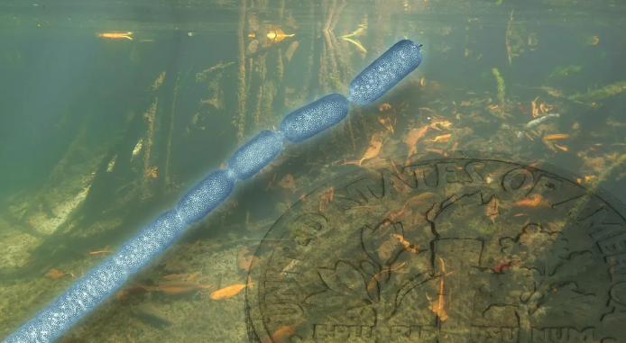
This unusual size is very noteworthy because bacteria are often invisible without the help of a microscope. "It's 5,000 times larger than most bacteria." Ean-Marie Volland said he is a scientist at the U.S. Department of Energy's (DOE) Joint Genome Institute (JGI), a user facility in the DOE Office of Science, based at the Lawrence Berkeley Lab and the Complex Systems Research Laboratory (LRC) in Menlo Park, California, with joint appointments. In the June 24, 2022 issue of the journal Science, Volland and colleagues, including researchers at JGI and Berkeley Labs, LRC, and the University of Guadeloupe, described the morphology and genomic characteristics of this giant filamentous bacterium, as well as its life cycle.
For most bacteria, their DNA floats freely in the cytoplasm of their cells. This newly discovered bacterial species makes its DNA more organized. "The biggest surprise of the project was the realization that these genomic copies that spread across the entire cell were actually contained in a membrane-like structure," Volland said. "And that's very unexpected for a bacterium."

Strange encounters in the mangroves
The bacteria themselves were discovered in 2009 by Olivier Gros, a professor of marine biology at the University of Antilles in Guadeloupe. Gros' research focuses on marine mangrove systems, and when he first encountered the bacteria, he was looking for symbiotes of sulfur oxide in sulfur-rich mangrove sediments not far from his lab. "When I saw them, I thought, 'Weird,'" he said. At first I thought it was just something curious, some white filament that needed to be attached to something in the sediment, like a leaf. Lab researchers did some microscopic studies over the next few years and realized that it was a prokaryote of sulfur oxide.
Silvina Gonzalez-Rizzo, associate professor of molecular biology at the University of Antilles and co-first author of the study, performed 16S rRNA gene sequencing to identify and classify this prokaryote. "I thought they were eukaryotes; I don't think they're bacteria because they're so big that there seem to be a lot of filaments," she recalls her first impressions. "We realized they were unique because it looked like a single cell. They are a 'macro' microbe, which is fascinating! ”
"She understood that this was a bacterium that belonged to the genus Thiomaragarita," Gros noted. "She named it Ca. Thiomargarita magnifica。 ”
"Magnifica is because magnus means big in Latin, and I think it's as gorgeous as the French word magnifique," Gonzalez-Rizzo explains. "This discovery opens up new questions about bacterial morphology that have never been studied before."
Characterize giant bacteria
When Volland returned to Gross's lab as a postdoctoral fellow, he was involved in the study of the giant Thiomaragarita bacteria. He will be working at JGI when he applies for a discovery position at LRC, and Gros allows him to continue his research on the project.
At JGI, Volland began working on Ca. T. magnifica was in Tanja Woyke's single-cell group to better understand the role of this sulfur oxide, carbon-fixed bacterium in mangroves. "Mangroves and their microbiomes are important ecosystems for the carbon cycle. If you look at the space they take up on a global scale, it's less than 1% of the world's coastal areas. But when you look at carbon storage again, they contribute 10-15 percent of the carbon stored in coastal sediments," said Woyke, who is also the director of the JGI Microbiology Project and one of the paper's senior authors. Given the potential interactions of these large bacteria with other microbes, the team also had to study them. Woyke said: "We started this project under the strategic thrust of JGI's biological interactions, as large sulfur bacteria have been shown to be hotspots for symbionts. She added: "However, this project has taken us in a very different direction. ”
Volland accepted the challenge of visualizing these giant cells in three dimensions at a relatively high magnification. For example, using various microscopy techniques, such as hard X-ray tomography, he saw entire filaments up to 9.66 millimeters long and confirmed that they were indeed giant single cells, not multicellular filaments, which are common in other large sulfur bacteria. He was also able to take advantage of Berkeley Lab's existing imaging facilities, such as confocal laser scanning microscopy and transmission electron microscopy (TEM), to look at filaments and cell membranes in greater detail. These techniques allowed him to observe new membrane-bound chambers containing DNA clusters. He called these organelles "pepins," named after the small seeds in the fruit. DNA clusters are very numerous in single cells.
The team understood the genome complexity of the cell. As Volland points out, "These bacteria contain three times more genes than most bacteria, as well as hundreds of thousands of copies of the genome (polyploidy) that are spread throughout the cell." The JGI team then analyzed five bacterial cells at the molecular level using single-cell genomics. They amplified, sequenced and assembled the genome. At the same time, Gros' lab also used a labeling technique called BONCAT to identify regions involved in protein-making activities, which confirmed that entire bacterial cells are active.
Shailesh Date, one of the article's senior authors and founder and CEO of LRC, said: "This project is a great opportunity to demonstrate how complexity has evolved in some of the simplest organisms. One of the things we are arguing for is that it is necessary to observe and study the complexity of living things in more detail than is currently being done. So we think very, very simple organisms may have some surprises. ”
LRC funded Volland through grants from the John Templeton Foundation and the Gordon and Betty Moore Foundation. Sara Bender of the Gordon and Betty-Moore Foundation added: "This groundbreaking discovery underscores the importance of supporting fundamental, creative research projects to advance our understanding of the natural world. We look forward to learning about Ca. We look forward to learning about Ca. How the characteristics of Thiomargarita magnifica challenge the current patterns that make up bacterial cells and advance microbial research. ”
A giant bacterium, multiple research questions
For the team, the description of Ca. Thiomargarita magnifica paves the way for several new research questions. Among them, the role of this bacterium in mangrove ecosystems. "We know it grows and thrives on sediments of mangrove ecosystems in the Caribbean," Volland said. Metabolically, it undergoes chemical synthesis, a process similar to plant photosynthesis. "Another open question is whether the new organelles, named pepins, played a role in the evolution of the extreme size of Thiomaragarita magnifica, and whether pepins were found in other bacterial species." The precise formation of pepins and how molecular processes inside and outside these structures occur and are regulated also needs to be studied.
Both Gonzalez-Rizzo and Woyke believe that successfully culturing these bacteria in the lab is a way to get some answers. "If we can sustain these bacteria in a lab environment, we can use technology that isn't feasible today," Woyke said. You can find some pictures of TEM and see things that look like pepins, so maybe people see them but don't understand what they are. It would be very interesting to check if pepins were already everywhere. ”