Today, when we walk into the hospital, we must feel that nuclear medicine technology is omnipresent: X-ray imaging, CT imaging, magnetic resonance imaging, various radiation therapy technologies, etc. escort our health, but also greatly promote the development of modern medicine.
The "magnetic resonance imaging" we usually hear is actually magnetic resonance imaging (MRI), also known as magnetic resonance imaging. Since 1937, when Isador Isaac Rabi discovered the phenomenon of mriori, magnetic resonance technology has developed rapidly in the decades that followed and is now an indispensable device for clinical examination and diagnosis. In the development of magnetic resonance technology, a total of 6 Nobel Prizes have been born.
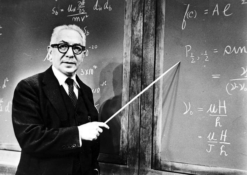
Figure 1 Isador Isaac Rabi (1898-1988) won the 1944 Nobel Prize for discovering the phenomenon of nuclear magnetic resonance. The source of the image is | researchgate

Figure 2 Development of MRI technology Figure | Yu Boyi
What is the photo of MRI? How do magnetic resonance devices get images of the structure of tissues in our bodies? We can understand it from the "nuclear", "magnetic", "resonance", "imaging" parts.
01
The "nucleus" of MRI
We know that the human body is made of atoms, and atoms contain atomic nuclei and extranuclear electrons. The "nucleus" of NMR is actually the nucleus of an atom. There are many types of nuclei, and can each of them be mrizuliographed? Of course not, only magnetic nuclei can be mriographed.
The magnetic moment of the nucleus is derived from the magnetic moment of the nucleus, and the magnetic moment of the nucleus is derived from the spin angle momentum of the nucleus. We can think of the nucleus as a sphere, and all magnetic nuclei have a characteristic, that is, they rotate at high speed around their own axis, and we call this property of the nucleus spin.
Simply put, the number of protons and neutrons in an atomic nucleus determines whether the nucleus is a magnetic nucleus or not. If the number of protons and neutrons in the nucleus is even, then such a nucleus cannot spin to produce nuclear magnetism, which is a non-magnetic nucleus, and vice versa.
Since the surface of the nucleus is positively charged, the spin of the magnetic nucleus produces a ring current, which produces a magnetization vector with a certain size and direction. We call this magnetic field generated by the spin of a magnetic nucleus nuclear magnetic.
Figure 3 We can think of an atomic nucleus as a sphere. The positive charge on the surface of the nucleus rotates at high speed to produce a ring current, which in turn produces a magnetization vector of a certain size and direction. Pictured| Li Jiaxin
Even so, there are still more magnetic nuclei in our bodies, what kind of magnetic nuclei are chosen for magnetic resonance imaging of our human body?
Table Common Magnetic Nuclei in The Human Body Data Sources| "Magnetic Resonance Imaging Technical Guidelines--- Examination Specifications, Clinical Strategies and New Technology Applications
As shown in the table above, hydrogen nuclei have the highest content in the human body, and the susceptibility is also the highest, so we generally use hydrogen nuclei for magnetic resonance imaging. Therefore, when you shoot NMR, you shoot the "hydrogen nucleus", which can also be called "proton" (because there is no neutron in the hydrogen nucleus, only a proton).
The hydrogen nucleus in the human body mainly comes from three types of compounds - water, fat, and protein, but the hydrogen nucleus in the protein generally does not have an MRI signal, so the MRI signal in the human body mainly comes from water, and the signal in some tissues also comes from fat.
02
The "magnetism" of MRI
Since the hydrogen nuclei in our bodies all have nuclear magnetism, and the hundreds of millions of hydrogen nuclei in each of our bodies produce magnetic fields, why haven't we become magneto?
Figure 4 Magneto is a Marvel Comics supervillain who can control any form of magnetic field. The source of the image is | Baidu Encyclopedia
This is because the hydrogen nuclei in our bodies are disordered, and the magnetization vectors in all directions cancel each other out, so macroscopically we are not magnetic.
So, how do you observe the magnetic resonance signal in the body?
Each hydrogen nucleus produces a magnetic field similar to a small magnetic needle that is deflected by a force in the magnetic field. If we add a main magnetic field, due to the force in the magnetic field, the magnetization vector produced by the hydrogen nucleus will be parallel to or parallel to the direction of the main magnetic field.
Protons parallel to the main magnetic field are at a low energy level without the need to resist the action of the main magnetic field; protons parallel to and reverse the main magnetic field need to be at a high energy level against the action of the main magnetic field. There are slightly more protons at the low energy level than protons at the high energy level, and the whole is macroscopically represented as a magnetization vector in the same direction as the main magnetic field.
Figure 5 A, the magnetization vector of each proton is in a different direction under the action of no magnetic field in vivo protons; B, when the human body is in the external magnetic field, under the action of the external magnetic field, the magnetization vector of the protons in the body will face in two directions. Figure | Yu Boyi
It's like a student training in the playground, and during intermission, everyone is doing things at random. However, when the instructor's password was heard, "Stand in two rows facing the sun and back to the sun," the students would immediately stand in two rows. Because they can't stand the harsh sunlight, more students instinctively stand in the row with their backs to the sun.
It should be noted that the hydrogen nucleus in the magnetic field is not completely parallel to the direction of the main magnetic field, but there is a certain angle. Therefore, in addition to spinning in the main magnetic field, the hydrogen nucleus rotates around the axis in the direction of the main magnetic field, moving in the same way as a tilted gyroscope on the ground. We call this movement the Larmor procession.
Figure 6 Spinner protons precede like gyroscopes in a magnetic field. Pictured| Li Jiaxin
The frequency ω of the Larmor precession can be described by the following formula:
Where γ is the magnetic rotation ratio of the atomic nucleus, which is generally a constant; B is the main magnetic field strength. The magnetic spin ratio of the hydrogen nucleus is 42.5MHz/T.
03
"Resonance" of MRI
When it comes to resonance, it's easy to think of the resonance of sounds you learned in junior high school. Similarly, the condition for sound resonance is that the sound waves are consistent with the vibrational frequency of the tuning fork, and similarly, the condition of NMR is that the applied rf pulse is consistent with the proton's Larmor precessive frequency.
As mentioned earlier, under the action of the magnetic field, the protons in vivo in the macroscopic area produce the same magnetization vector (Mz) as the main magnetic field, and the microscopic number of protons in different energy levels is different, and the splitting of this energy level will only occur when the magnetic field exists.
Just like giving a student with his back to the sun, so that he can also stand in the row facing the sun, when we apply an RF pulse that is consistent with the proton precession frequency, the low-energy protons will resonate, absorb the RF energy and jump to the high-energy level, and the number of protons at the high-energy level will gradually increase.
When the number of high-energy protons and low-energy protons coincides, and the magnetization vector cancels out each other, the magnetization vector of the proton in the direction of the main magnetic field is 0 at the macroscopic level (as shown in the lower left side of Figure 7).
But at the same time, due to the convergence effect of radio frequency, the magnetic vector in the XY direction will be gathered together to produce a macroscopic magnetization vector of the XY plane, rotating around the magnetic field. This polyphase effect, just as our five fingers are originally open, the five fingers are in all directions, if each finger represents a force, the combined force is zero; when we close the fingers, the direction of the combined force is the direction in which the five fingers converge. (As shown below on the right side of Figure 7)
Figure 7 Under the action of the magnetic field, the hydrogen atom splits at the nuclear level and undergoes nuclear magnetic resonance under the action of the radio frequency pulse, resulting in an energy level transition. Figure | Yu Boyi
04
"Imaging" of MRI
When we remove the RF pulse, the protons at the high energy level will gradually return to the low energy level. Macroscopically, it is manifested as the gradual recovery of the longitudinal magnetization vector (longitudinal relaxation, Fig. 8) and the gradual decay of the transverse magnetization vector (transverse relaxation, Figure 9).
Fig. 8 Gradual recovery of longitudinal magnetization vector Fig. | Li Jiaxin
Figure 9 Gradual decay of the lateral magnetization vector Figure | Li Jiaxin
Due to the different number of hydrogen nuclei in the human body tissue, the chemical environment in which the hydrogen nucleus is located is different, so the process of longitudinal and transverse relaxation is not the same, and the corresponding time for the recovery of the magnetization vector is also different.
Therefore, by setting up the MRI sequence, we can acquire the signal of the tissue. With different mri signal intensities of different tissues, we can get a comparative image of the tissues in vivo.
How do we acquire magnetic resonance signals? In fact, it is very simple, high school physics class tells us that "magnetic induction wire cutting coil will produce current", therefore, with the rotation of the XY direction of magnetization vector to cut the coil can get the electrical signal generated by the magnetic resonance, and then use mathematical methods to convert the electrical signal, you can obtain a magnetic resonance image.
The author | Yu Boyi Li Jiaxin
The instructor | Chen Weiqiang
Edit the | Liu fang
bibliography:
[1] Li Donglin,Gao Jiahong. Nuclear Magnetic Resonance Imaging—Physical Principles and Methods. Beijing:Peking University Press, 2014. 9.
[2] YANG Zhenghan,FENG Feng,WANG Xiaoying. Magnetic Resonance Imaging Technical Guidelines: Examination Specifications, Clinical Strategies and Application of New Technologies. Beijing:People's Military Medical Publishing House, 2010.
[3] Breneman, B. History, Physics, and Design of Superconducting Magnets for MRI. Emagres 8, 137-156.
[4] Collins, J. The history of MRI. Semin. Roentgenology 43, 259-260.
The reproduced content represents the views of the author only
Does not represent the position of the Institute of Physics, Chinese Academy of Sciences
If you need to reprint, please contact the original public account
Source: Institute of Modern Physics, Chinese Academy of Sciences
Edit: Cloud open leaves fall