For X-rays, CT, ultrasound, MRI, these common imaging tests, many people are not clear. Now, we compare the body to food and show you how these tests play a different role.
X-ray
It's like squashing the bread to look at
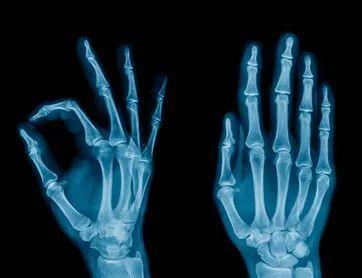
X-rays will pass through the human body, encounter the part that is obscured, the negative will not be exposed, and the part will be white after washing.
Like a slice of bread or a piece of cotton, you can't see the texture of the fibers inside, but it will be clearer when you press it by hand. The biggest disadvantage of x-rays is that images subject to depth and shallow tissue overlap and hide from each other, and sometimes multiple multi-angle X-rays are required.
CT
Like taking a slice of bread to see

The principle of CT examination is that the X-ray will pass through the human body in layers, and then the second imaging will be calculated by the computer, just like cutting a slice of bread into slices. The advantage is that it can be viewed hierarchically, and more organizational information can be displayed after calculation.
B-ultrasound
Like picking a watermelon before knocking
The principle of B ultrasound is to penetrate the human body with ultrasonic waves, and when sound waves encounter human tissue, reflected waves are generated and imaged by calculating reflected waves. It's like picking a watermelon, knocking on the side to see the lesions.
nuclear magnetic resonance
Shake it and look again
The nuclear magnetic resonance machine uses a more powerful magnetic field to make the magnetic field line of all water molecules in the human body consistent, when the magnetic field of the magnetic resonance machine suddenly disappears, and the magnetic field direction of the water molecules in the body suddenly returns to the original state of random arrangement. Simply put, it is equivalent to shaking it with your hand, letting the water molecules vibrate, and then calm down and feel the vibration inside. Therefore, MRI is also jokingly described as a shaky look.
What are the four methods used for?
At the time of consultation, doctors often prescribe a variety of imaging tests: ultrasound, CT, MRI... Many patients will question the doctor's deliberate high-priced examination order. In fact, doctors choose different imaging tests according to different conditions.
1
Traumatic bone - look at X-rays, look closely at CT, MRI can't see clearly
All kinds of trauma, if you suspect that the bone is injured, X-ray photos are preferred, and the examination results are quick and easy to obtain. For further observation, CT can be selected. Ultrasound, MRI is not very clear for the bone cortex medulla, etc., and generally do not choose.
2
Cervical and lumbar spine - the best MRI selection, followed by CT
Cervical spondylosis, lumbar disc herniation and other disc diseases need to observe the disc and the corresponding nerve roots, in order to better observe these soft tissues, the best choice is mrimagnetism. Similarly, for joint, muscle, and adipose tissue examinations, MRI is also preferred.
3
Chest - look at the X-ray roughly, look closely at the CT, look at the lungs and don't choose MRI
Chest x-ray can roughly examine the heart, aorta, lungs, pleura, ribs, etc., and can check for increased lung texture, calcification points in the lungs, calcification of the aortic nodes, etc.
Chest CT shows a clearer structure, and the sensitivity to chest lesion detection and the accuracy of showing lesions are better than conventional chest x-ray chest x-ray, especially for the early diagnosis of lung cancer. However, the radiation dose for CT examination is higher than the X-ray. The use of MRI in the diagnosis of lung disease is very limited.
4
Abdominal pelvis – except for the intestine, it can generally be examined by ultrasound
The abdominal organs are greatly affected by breathing, which in turn affects CT and MRI, and ultrasound is not affected. At the same time, ultrasound has a high accuracy in diagnosing organs such as liver, spleen, pancreas, kidneys, and pelvics.
However, ultrasound is greatly disturbed by gas, and for organs with more gas content such as the intestine, the accuracy of ultrasound diagnosis will be reduced.
5
Heart - CT for coronary heart disease, ultrasound for cardiac function
Routine heart structure and function examination, the information provided by cardiac ultrasound has been sufficient, and it is simple and easy to implement.
CT can examine the coronary arteries, but coronary CT examination has a large amount of radiation and is not suitable for routine physical examination. Although MRI has no electromagnetic radiation, the coronary arteries are not as well observed as CT. Cardiac MRI is the "gold standard" for evaluating the structure and function of the heart.
Source: Health Times, Qingdao Daily