Red Net Moment December 17 news (correspondent Lan Shujie Chen Zhiwu) Recently, the radiology team of Zhuzhou People's Hospital successfully carried out cranial CT perfusion imaging technology (CTP), which provided a more reliable basis for the clear diagnosis and scientific treatment of patients in the stroke center of the hospital.
With the improvement of people's living standards, more and more patients with stroke disease, the age of onset is getting younger and younger, and it has become the most common cerebrovascular disease. In clinical applications, due to the limitations of routine CT examination, the lesions of cerebral infarction found at this stage of examination, the damage to brain tissue has become irreversible, and valuable time for thrombolytic interventional surgery has been missed. How to detect lesions early, diagnose and treat early has become the most concerned clinical issue.
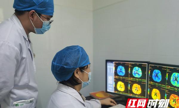
Cranial CT perfusion imaging can help patients more accurately assess the degree of brain tissue damage, in the rapid intravenous mass injection of contrast agent, through the continuous repetition of multiple times of rapid dynamic scanning of the region of interest, so as to obtain cerebral blood flow, blood flow speed, average passage time of blood flow, maximum peak time and other hemodynamic parameters and perfusion image performance to evaluate the perfusion status of brain tissue, used to assess brain tissue function. CTA (CT vascular imaging) combined with CTP can not only show the morphology, malformations, stenosis, etc. of blood vessels from the appearance and morphology, but also conduct qualitative and quantitative analysis of the infarction area. I believe that most people have had the experience of infusion, blood vessels are equivalent to our infusion tubes, CTA technology can intuitively see the external form of the infusion tube and the flow of liquid, and CTP can focus on observing the abnormal flow of fluid in the infusion tube on the basis of CTA qualitative and quantitative assessment. This technology provides reliable imaging data for whether thrombolysis can be achieved, which has the advantages of relatively non-invasive, convenient and fast, efficient and accurate, and is an important means for the early diagnosis and evaluation of staging of clinical ischemic stroke.
"Accurate imagery, technology first". The development of CT whole brain perfusion imaging technology marks that the basic imaging examination required by the stroke center of the hospital has been configured, which has far-reaching significance for the control of the disease of patients with acute stroke and chronic narrow occlusive vascular disease, and can enable more patients with ischemic cerebrovascular disease to get early diagnosis and treatment, and more patients with ischemic cerebrovascular disease to get early diagnosis and treatment.
![What are the chances of recovery after cerebral infarction? Doctor: It has a lot to do with these 5 factors![fig]](data:image/gif;base64,R0lGODlhAQABAIAAAP///wAAACwAAAAAAQABAAACAkQBADs=)