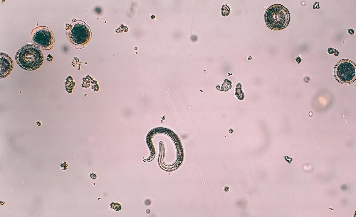
In recent years, people's living habits, environmental changes and the rise of pet fever have made parasitic infections extremely common in China's population. Some studies have found that women of childbearing age can cause various gynecological diseases after infection with parasites, but because patients have no obvious signs and symptoms after parasitic infection, coupled with backward detection methods, gynecologists generally do not consider the relationship with parasitic infections when diagnosing gynecological diseases, so they often cause misdiagnosis or missed diagnosis, which is very harmful to women.

<h1 class="pgc-h-arrow-right" data-track="25" > Toxoplasma gondii</h1>
Toxoplasma gondii, also known as toxoplasma gondii and toxoplasma gondii, is a parasitic protozoa in cells, which mainly causes damage to tissues and organs, and the reproductive system is no exception. Toxoplasma gondii is distributed around the world, 1/4 of the global population is threatened, and the infection rate of pregnant women toxoplasmosis is as high as 6% to 33%, and there is a trend of increasing year by year.
Toxoplasma gondii is transmitted vertically by placental mode, which is the cause of congenital toxoplasmosis and the most harmful mode of transmission of toxoplasma gondii to humans; transmission through the digestive tract and skin, such as drinking raw milk or eating raw meat, semi-raw meat, often contact with cats, dogs and other animals, is the most common route of transmission. Transmitted through blood, this mode of transmission is an important route for iatrogenic diseases, such as hemorrhagic shock, organ transplantation and other patients.
When Toxoplasma gondii infects the reproductive system of a person or animal, it will enlarge or dysfunction the endometrium and muscle layer, causing an inflammatory response to tissues such as the pelvic cavity, endometrium, and fallopian tubes. If there is diencephalonia damage after toxoplasma gondii infection, neuroendocrine disorders can occur in women, resulting in pelvic inflammatory disease, functional uterine bleeding, infertility, ectopic pregnancy, etc. In addition, the recent rate of infection with uterine fibroids, ovarian cysts, endometriosis, and ovarian teratomas is also higher than in women who are not infected with Toxoplasma.
Toxoplasma gondii has the characteristics of the central nervous system, mainly invades human brain tissue, and has the most suitable conditions for its survival in brain tissue. Therefore, Toxoplasma gondii is also considered to be the first of the five major pathogens caused by intrauterine infection in pregnant women that lead to embryonic malformations.
If a woman is detected toxoplasmosis during pregnancy, it can lead to adverse outcomes such as miscarriage, stillbirth, or teratology in the pregnant woman. However, not all fetuses infected with Toxoplasma gondii in the womb are stillborn or die immediately after childbirth, and among the fetuses born to pregnant women, there are malformed or congenital defects, and there are congenital Toxoplasma gondii infections of newborns with no abnormalities. However, as newborns grow older, they will have significant sequelae in their childhood or adolescence (about 85%), mainly eye and central nervous system damage, such as retinopathy, hydrocephalus, intracranial calcification, microcephaly, psychomotor disorders and even epilepsy.
Toxoplasma gondii first causes direct damage to embryonic cells. After Toxoplasma go through the blood to infect the placenta, it can pass through the placental barrier and directly infiltrate embryonic cells and tissues. It parasitizes within the cell and multiplies rapidly until the cell is destroyed, and at the same time, it invades adjacent tissues, so repeated destruction can cause cell and tissue defects and inflammatory reactions of the impregnated tissues. Second, Toxoplasma gondii can cause the absolute number of cells in the embryo to decrease, affect cell proliferation, and delay the intrauterine development of the fetus; in addition, Toxoplasma gondii can cause changes in chromosomal structure. Toxoplasma gondii in the early stage of embryonic development mainly destroys embryonic cells, causing the phenomenon of division and non-separation during the process of egg cleavage, resulting in chromosomal aberrations, resulting in the occurrence of malformations.
Although pregnant women infected with Toxoplasma gondii can endanger embryos and fetuses, in real life, not all infected pregnant women will spread to the fetus, and only a few pregnant women will have intrauterine transmission. Whether a pregnant woman will cause fetal malformations after infection and the severity of the cause are very related to these factors: (1) The period of infection in pregnant women. Studies have shown that the incidence of Toxoplasma gondii infection will gradually increase with the progression of early, middle and late pregnancy, but the degree of harm to the fetus will gradually decrease. Among them, the first trimester of pregnancy causes the greatest damage to the embryo, which can easily lead to miscarriage, stillbirth, and severe multiple malformations. (2) Virulence of insect strains: strong virulence can quickly cross the placenta and blood-cerebrospinal fluid barrier, causing the spread and vertical spread of pathogens before the body produces effective immune defenses. (3) The body's immunity. Studies have found that previous chronic infections in the mother protectively block vertical transmission during re-infection during pregnancy. In mothers in the first trimester, high titers of IgG are important markers of resistance to toxoplasma gondii primary infection, reducing the risk of fetal infection in the mother.
<h1 class="pgc-h-arrow-right" data-track="24" > Amoeba</h1>
Amoebic disease is a more aggressive intrahistolytic amoebic infection. It spreads naturally between humans and animals, and in general, amoeba parasitizes in the large intestine and sometimes appears in the intestinal mucosa or other organs. Among patients infected with amoebic protozoa, only 10% of infected patients will have clinical symptoms, such as amoebic bowel disease, amoebic liver abscess, etc. Cutaneous amoeba disease can invade the human cervical, vaginal and urethral reproductive systems.
Intrahistolytic amoebic genital tractitis is an inflammation of the reproductive tract caused by amoebic protozoa infection, mainly caused by intestinal infections. Usually, the amoebic trophozoite in the patient's stool is excreted with fecal mucus and can spread directly to the vulva and vaginal opening, but in healthy conditions or washed away by urine, or uninfected because the vaginal mucosa is intact. If the health level of the body declines, the general condition is poor, or the reproductive tract is damaged, Amoebic protozoa can take the opportunity to invade the reproductive tract tissue, dissolve and destroy the reproductive tract tissue, so that the vaginal mucosa necrosis, the formation of marginal bulging, irregular, granular shallow ulcers, the surface of the ulcer is often covered by yellow-brown necrotic tissue, there are purulent or bloody secretions discharged. Most gynecologists are prone to misdiagnosis of vaginal cancer due to lack of knowledge about tissue-lysolytic amoebic reproductive tractitis. The most reliable diagnostic method is to find amoeba trophozoites from vaginal secretions. In patients with chronic ulcers with negative discharge, a biopsy should be done to correctly identify the cause.
<h1 class="pgc-h-arrow-right" data-track="23" > trichomoniasis vaginalis</h1>
Trichomonas vaginalis infection is one of the most prevalent infectious diseases of the reproductive tract and one of the most prevalent sexually transmitted diseases. More than 200 million people worldwide are infected with Trichomoniasis vaginalis each year. Almost all foreign studies have shown that Trichomoniasis vaginalis is transmitted entirely through sexual activity, and only individual reports have other indirect transmission routes. Therefore, the World Health Organization has long included Trichomoniasis vaginalis in the scope of sexually transmitted diseases.
Trichomonas vaginalis was first discovered in 1836 from purulent vaginal secretions and male genitourinary secretions, and the latest data show that after infection with trichomonas vaginalis, the susceptibility to other sexually transmitted disease pathogens can also increase the susceptibility to other sexually transmitted disease pathogens, which can increase the chance of infection with immunodeficiency virus (HIV) by 2 to 4 times.
The vaginal environment of healthy women can inhibit the growth and reproduction of insects or bacteria, which is what we usually call the self-purification of the vagina. However, Trichomonas vaginalis can destroy the "vaginal self-purification effect", so that Trichomonas vaginalis can multiply in large quantities, and then bacterial infections occur, resulting in inflammatory lesions of the vaginal mucosa.
After infection with trichomonas vaginalis, women mainly cause trichomoniasis vaginitis and urethritis, which can invade the cervix, paraurethral glands, bladder, and even fallopian tubes. The typical symptom of trichomoniasis vaginitis is increased vaginal discharge, a thin foamy white, yellow, or green color, itching of the vulva, and if there is a mixed infection of other bacteria, the vaginal discharge is purulent and odorous. The itching site is mainly concentrated in the external vaginal opening and vulva, and sometimes there are symptoms such as burning, pain, and dyspareunia. Chronic trichomoniasis vaginalis infection may have only white, yellow, and green vaginal discharges without other symptoms. Some of the female vaginal trichomoniasis infections have no symptoms and become carriers. In addition, vaginal trichomoniasis infection is closely related to the incidence of cervical cancer; trichomonas vaginalis also has the effect of devouring sperm, which can lead to infertility in women and can increase the chances of HIV infection. Infection of pregnant women with trichomonas vaginalis can induce preterm birth, stillbirth, and birth of low-weight infants.
Trichomoniasis vaginalis has a strong ability to live, and if patients cannot receive correct and effective treatment, they are prone to recurrence. The correct method of medication is to stuff the tablets into the top of the vagina, so that the tablets can evenly contact the vaginal wall after melting, which plays a killing role. Do not put the pill in the vaginal opening, or get up and move or urinate shortly after putting it in, causing the pill to fall out and lose its therapeutic effect. At the same time, when the wife suffers from trichomoniasis vaginitis, due to the contact of sexual life, the husband's urinary tract will also be infected with trichomoniasis, it is best to treat both parties at the same time to avoid repeated infection, long-term incurable phenomenon.
<h1 class="pgc-h-arrow-right" data-track="19" > hydatid disease</h1>
Echinococcosis is a common parasitic disease that occurs mostly in the northwest region of China, mainly in organs such as the liver, lungs and abdominal cavity, and there are not many violations of the female reproductive tract. After a person swallows food contaminated with eggs, its larvae six hooked larvae enter the bloodstream and spread through the left heart to distant organs throughout the body, and the degree of infection of each organ is very related to the blood flow in the arteries. Due to endocrine relationships, blood flow to the reproductive organs and pelvis increases during menstruation and pregnancy, and the chance of echinococcosis infection with blood flow also increases. In addition, echinococcosis eggs can be implanted directly under the vaginal mucosa, and under the action of vaginal secretions, they gradually develop into echinococcosis cysts.
Echinococcosis of the genital tract does not affect menstruation and pregnancy, some patients' echinococcosis cysts will compress adjacent organs, appear pain symptoms, and the vast majority of echinococcus growing in the pelvic cavity and the large omentum, peritoneum, reproductive organs adhesion, the lump wall is relatively thick, the texture is also relatively hard, easy to be misdiagnosed by doctors is ovarian tumor or teratoma.
Parasitic infections can cause a variety of gynecological diseases, and the clinical symptoms are more complex, easy to misdiagnosis, seriously affecting women's physical health, worthy of great attention. Common points in the prevention of parasitic infections: establish good living habits and hygiene habits, especially women of childbearing age should avoid frequent contact with animals or domestic pets, minimize the number of meals outside, do not eat raw meat, etc. In addition, pay attention to personal hygiene, especially pay attention to menstrual hygiene and sexual intercourse hygiene, and underwear should be washed in a basin with other clothing.