The caudate lobe (segment I) has a separate venous return system, which is the only posterior segment of the posterior hepatic segment in which venous blood is injected directly into the inferior vena cava through a series of short hepatic veins, herein the third hepatic hilar.
In addition, the hepatic caudate lobe also has a separate drainage bile duct, and at the same time by the left and right hemispheres and hepatic artery branches to supply blood, based on this special anatomical structure, the hepatic caudate lobe has extremely important clinical significance.
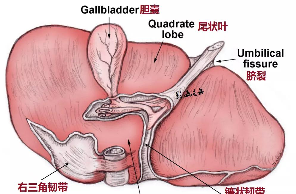
Note: umbilical fissure translates to umbilical fissure, but the word seems to have not heard much since school, checked the relevant information, found that this umbilical fissure is the hepatic round ligament fissure.

By the way, review the liver lobular anatomy~
In patients with early Budd-Gart syndrome (30-year-old women), the volume of the hepatic caudate lobe (cl) is significantly increased, and the rest of the liver parenchyma contour is acceptable, but the degree of strengthening is significantly reduced, and it is scattered in patchy intensification (white arrow), which is caused by hepatic congestion, and the effusion (asterisk) can be seen in the abdominal cavity, and the triangle refers to the inferior vena cava of the non-affected segment.
The hepatic caudate lobe can be "left alone" in Budd-Gart syndrome, thanks entirely to its unique anatomy. Most patients with cirrhosis may also see enlarged caudate lobes.
In patients with advanced Buddart syndrome (inferior vena cava diaphragm), the caudate lobe of the liver is still compensated for enlargement, local strengthening is relatively uniform, the rest of the liver parenchyma is unevenly strengthened, irregular fissures are seen without strengthening areas, and the liver envelope is not polished, showing compensatory cirrhosis. Note that collateral angiogenesis (yellow arrow) is visible on the right side of the spine, and there is no significant effusion in the abdominal cavity.
It has also been reported in the literature that cholangiocarcinoma of the hepatic caudate lobe does not produce signs of intrahepatic bile duct obstruction, which has an independent drainage of the bile ducts and has a similar effect to intravenous drainage.
Source: Shadow Sea Rafting
【Copyright Notice】This platform is a public welfare learning platform, reprinted for the purpose of transmitting more learning information, and has indicated the author and source, such as teachers who do not want to be disseminated can contact us to delete