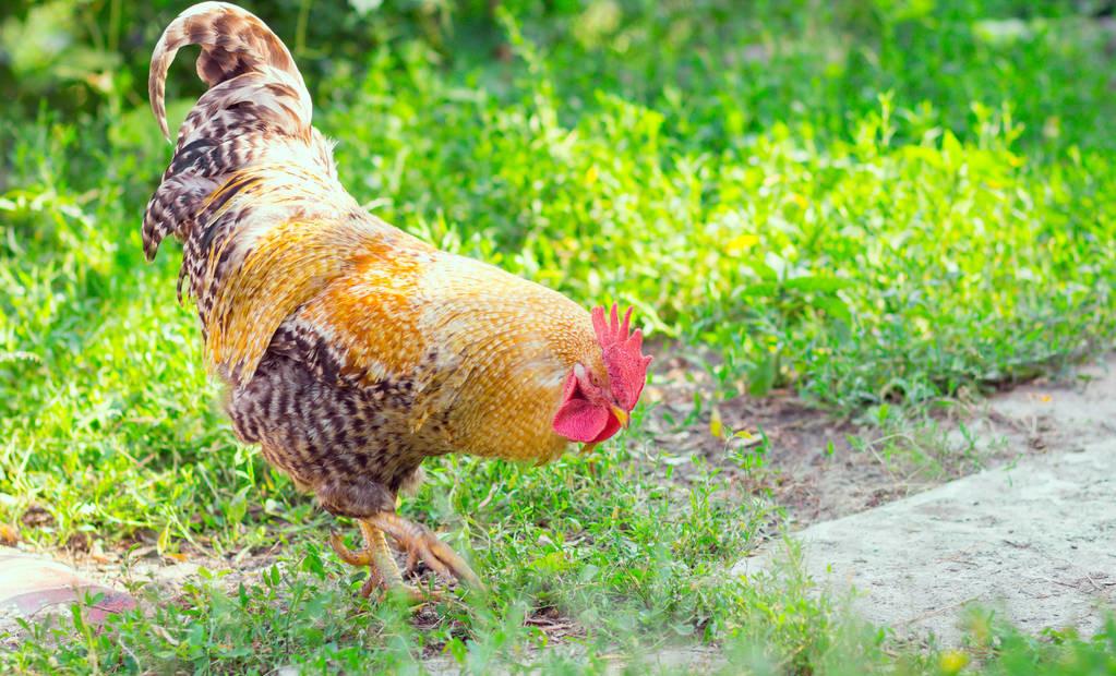
First, the reproductive system of chicken males
The reproductive system of a rooster consists of the testicles, epididymis, vas deferens, and mating organs.
The testicles of the rooster are paired, bean-shaped, located in the abdominal cavity, suspended in a short mesenteric over the ventral side of the anterior lobe of the kidney, surrounded by abdominal balloons. The testicles of small roosters are small, such as the size of rice grains, yellow; the adults are significantly enlarged, and the testicles are the largest in the breeding season after sexual maturity, such as the size of a pigeon's egg, yellowish white or white. The rooster's semen is weakly alkaline, with less ejaculation per ejaculation, but a high sperm concentration. Roosters begin to produce sperm at 12 weeks of age, and do not produce semen with a higher fertilization rate until 22 to 26 weeks of age, and roosters aged 1 to 2 years have the best semen quality.

The epididymis is attached to the dorsal medial border of the testicles and has the effect of storing sperm and secreting sperm. A vas deferens is a pair of curved tubes that secrete sperm, store sperm, and transport semen. The mating organs of the rooster are 3 small juxtapositions, called the body of the penis, located on the medial side of the ventral lip of the cloaca, hidden in the cloaca when not extended for mating, and erect and protruding during mating.
2. Chicken female reproductive system
The reproductive system of the hen consists of the ovaries and fallopian tubes, with only the ovaries and fallopian tubes on the left developing normally, and the right side developing stagnant and gradually degenerating during early embryonic development.
The ovaries are located in the left abdominal cavity, below the anterior lobe of the left kidney, and hang in a short mesary membrane of the lumbar dorsal side wall. The ovaries of young chickens are small, flattened oval, yellow-white or white, and the surface is granular; with the growth of age and sexual activity, the follicles gradually mature, due to the accumulation of a large amount of yolk in the follicle, protruding from the surface of the ovary, until the ovulation, only the follicle stalk is connected to the ovary, so the follicle is grape bunch-shaped; into the egg laying period, the follicle grows rapidly, the ovary common 4 to 5 larger follicles, the largest yolk-filled follicle diameter can reach 4 cm. At ovulation, the follicular membrane ruptures at a thin bloodless follicular spot, and the egg is released, the follicle has no follicular cavity and follicular fluid, and no corpus luteum is formed after discharge. When production is discontinued, the ovaries return to their shape and size at rest.
The fallopian tube is a long, curved tube that is 60 to 70 cm long during the egg laying period, retracted to 30 cm during the incubation period, and only 18 cm during the moulting period. The fallopian tubes can be divided into five parts from front to back, namely the funnel part, the protein secretion department (the large part of the bulge), the isthmus, the uterus and the vagina. Located behind the ovary, the funnel is the site of egg ingestion and fertilization, with free mucosal folds on its edges, called tubal umbrellas, and an abdominal opening of the fallopian tubes in the center. Protein secretion is long and curved, 30 to 50 cm long, the diameter of the tube is large, the wall of the tube is thick, and the dense glandular tube has the effect of secreting protein. The isthmus is short and narrow, 8 to 10 cm long, and there are glands in the mucous membrane that secrete keratin and form the inner and outer egg shell membranes. The uterus is pouch-shaped, with thick walls and about 10 cm long. There are shell glands on the mucosa of the uterus that secrete calcium, keratin and pigments to form eggshells. The vagina is the end of the fallopian tube, which opens to the left side of the dorsal wall of the cloaca, the mucous membrane of the vagina is white, forming a wrinkle, which can store sperm; the mucous membrane has glands, and its secretions form a thin layer of cornea on the surface of the egg shell.