<h1 class="ql-align-center">
Title II Parasites</h1>
Chapter II: Parasitic Diseases of Cattle and Sheep</h1>
<h1>
Section 1 Helminthiasis</h1>
First, trematodiasis
Trematodes refer to the bodies of the phylum Oblates, the family Trematodons. There are many species of trematodes parasitic in cattle and sheep, and the more serious ones in China are: Fasciola hepatica of the flaky family Flaky family, Dicrocoelium lanceatum of the genus Dicrocoelium lanceatum of the genus Dicrocoelium and Eurytrema pancreaticum of the genus Broad-plated, and Schistosoma japonicum of the genus Schistosoma japonicum. This type of disease has a slow onset and is characterized by digestive disorders.
cause of disease
1. Liver flaky trematodes The body of the insect is flattened leaves, the living body is tan, and when fixed, it is grayish white. It is 21 to 41 mm long and 9 to 14 mm wide. The anterior end of the worm has a triangular cone-like protrusion with a broader "shoulder" at the base, which gradually narrows from the shoulder to the back. The mouth suction cup is located at the front of the tapered protrusion, and the ventral suction cup is slightly larger than the mouth suction cup, just behind the center of the shoulder horizontal line. The genital foramen are between the mouth suction cup and the abdominal suction cup. The eggs are oblong-oval in shape, 133 to 157 μm in size× 74 to 91 μm in size, yellowish brown, with an unstard egg cap at the narrow end, and the egg is filled with yolk cells and an oocyte.
Also in the genus hepatic flaky trematodes are large-scale flukes (F. spp.). gigantica), the adult is 25 to 75 mm long, willow-leaf-shaped, without obvious double "shoulders", and the sides of the worm body are relatively straight. The eggs are yellowish brown, oblong and round, with a size of 150 to 190 μm × 70 to 90 μm.
2. Spear-shaped bilumen fluke Spear-shaped biluminal flukes are also known as bilocclus twigs (D. dendriticum), the insect body is flattened, narrow and "spear-shaped", the living body is brownish red, and it is off-white after fixation. It is 6.7 to 8.3 mm long and 1.6 to 2.2 mm wide. The mouth suction cup is located at the front end, and the ventral suction cup is located 1/5 of the front of the body. The digestive system consists of the mouth, pharynx, esophagus, and two simple intestinal tubes. 2 round or marginally missing testicles, anterior or posterior or diagonally behind the abdominal suction cups, and the male stem sac is located between the intestinal bifurcation and the ventral suction cups. The genital foramen is opened at the fork of the intestine. The ovaries are round and located behind the testicles. The yolk glands are small granular on either side of the middle of the worm body. The uterus is curved and fills the posterior half of the worm's body. The eggs are ovoid, yellowish brown, with an egg cap at one end, asymmetrical left and right, and contain hairy worms. The egg size is 34 to 44 μm× 29 to 33 μm.
Chinese double-lumen flux (D. chinensis) is of the same genus as spear-shaped bilobite flukes and has a similar morphology, but the body is wider, 3.5 to 9 mm long and 2 to 3 mm wide. The main difference is that the two testicles are edged or slightly lobed, juxtaposed left and right behind the abdominal suction cups.

3. Pancreatic broad-plated trematodes The body of the insect is flattened, oblong-oval, and the living body is brownish-red, and it is grayish-white after fixation. It is 8 to 16 mm long and 5 to 5.8 mm wide. The suction cups are well developed, and the mouth suction cups are significantly larger than the abdominal suction cups. The pharynx is small, the esophagus is short, and the two intestinal branches are simple. 2 testicles, round or slightly lobed, arranged left and right behind the ventral suckers. The male stem sac is tubular in the shape of a long tube, located between the ventral suction cup and the bifurcation of the intestinal branch. The ovaries are divided into 3 to 6 lobes, located behind the testicles. The fertilized vesicle is rounded and close to the ovaries. The uterus has many curves, is located in the posterior half of the worm body, and is filled with brown worm eggs. The yolk glands are granular and are located on either side of the central part of the worm body. The eggs are yellowish brown or tan, oval in shape, slightly asymmetrical on the sides, with an egg cap that contains an oval hairy lice. The egg size is 42 to 50 μm × 26 to 33 μm. Also in this genus are the E. coelomaticum and the E. cladorchis.
4. Japanese schistosomiasis Hermaphroditic, linear. The male is milky white, 10 to 20 mm in size× 0.5 to 0.55 mm in size, with a suction cup at the front of the body and a ventral suction cup behind it, with a short, thick stalk connected to the worm body. From behind the ventral suction cup to the tail, the sides of the body wall are rolled up to the ventral surface to form a cuddling groove, in which the female often resides, and the two are in a hugging state. The digestive organs have an orifice, an occlusal tract, and a lack of pharynx, and two intestinal tubes merge into one at 1/3 behind the worm body from before the abdominal suction cup. The male has 7 testicles, oval in shape, arranged in a single row behind the abdominal suction cup. The opening of the genital foramen is in the female groove behind the abdominal suction cup.
The female is dark brown in size from 15 to 26 mm × 0.3 mm, and is slenderr than the male. The mouth and abdominal suction cups are smaller than those of males. The ovary is oval in shape and is located between the two intestinal tubes in the middle of the worm body. The fallopian tubes fold forward and merge with the yolk tubes in front of the ovaries to form an egg mold. The uterus is tubular, located in front of the egg mold, and contains 50 to 300 eggs. The yolk glands are regularly branched and are located 1/4 behind the worm body. The genital foramen opens behind the ventral suction cups.
The eggs are oval in shape, pale yellow, with a thin, uncovered shell with a small spine on its side that contains trichoceratum. The egg size is 70 to 100 μm × 50 to 65 μm.
Developmental history
1. Liver flaky trematodes Adult worms parasitize in the liver bile ducts of cattle and sheep, the eggs laid enter the intestinal cavity with the bile, and then excrete with feces, under suitable conditions, the caterpillars are hatched and swim in the water after 10 to 25 days, and encounter the appropriate intermediate host - vertebral snails into it to develop into tail cercaria. Cysticers leave the snail body and shed their tails off aquatic plants or under the surface of the water to form cysticers. Cattle and sheep are infected by swallowing cysticers while eating grass or drinking water. Cysticercles escape from the duodenum, and it takes about 2 to 3 months for the child to mature through the intestinal wall and drill into the bile ducts in the liver through the surface of the liver.
2. Spear-shaped bilobites Adult worms lay eggs in the bile ducts and gallbladders of ruminants such as cattle and sheep, and the eggs enter the intestine with the bile and are excreted with feces. After being ingested by the first intermediate host, the land snail, the eggs develop into tail ceratolls in their bodies. A large number of tailed larvae gather to form a group of tail cymbs, which are wrapped in a sticky substance to form a sticky ball, which is discharged from the respiratory cavity of the snail, adheres to the plant leaves and other objects, and is swallowed by the second intermediate host, ants, and soon forms cysts in its body. The terminal host, cattle and sheep, is infected by swallowing ants containing cysticercles. Cysticers are desacialized in the intestines of cattle and sheep, and develop into adults from the duodenum through the total bile ducts and into the gallbladder for 72 to 85 days.
3. Pancreatic broad-plated trematodes Adult worms lay eggs in the pancreatic ducts of ruminants such as cattle and sheep, and the eggs enter the intestine with the pancreatic juice, and then excrete with feces, and are swallowed by the first intermediate host - land snails, and develop into larvae in their bodies. Mature larvae contain many cymbids, and the viscous mass of the larvae escapes from the snail. The second intermediate host, the grass lice, swallows the tail cymbal and develops into a cysticer in its body. Ruminants such as cattle and sheep become infected by swallowing grass liceps containing cysticers. After the cysticer is decanted from the duodenum of cattle and sheep, it enters the pancreatic tube from the opening of the pancreatic duct and develops into an adult worm after about 3 months.
4. Japanese schistosomiasis This insect is often referred to as schistosomiasis. Adults parasitize the portal vein and mesenteric vein of cattle and sheep, and after the female and males mate, the eggs produced by the females are partly flowing down the bloodstream to the liver, and some are piled up on the intestinal wall to form nodules. Eggs on the intestinal wall, after maturity, the eggs secrete lysozyme from the micropores of the egg shell to penetrate into the tissues, destroy the blood vessel wall, and cause inflammation and necrosis of the surrounding intestinal mucosal tissue, and at the same time, with the help of intestinal wall muscle contraction, so that the nodules and necrotic tissues break into the intestinal lumen, so that the eggs enter the intestinal cavity and are excreted with feces. The eggs fall into the water and quickly hatch under suitable conditions, which swim in the water and encounter the intermediate host, the snail, which burrows into its body and develops into the tail cercaria. The tail clyps leaves the snail body and swims on the surface of the water, encounters cattle and sheep, invades from the skin, and develops into adult worms through the right heart, lungs and systemic circulation through the blood flow through the right heart, lungs and systemic circulation through the small blood vessels and lymphatic vessels to the mesenteric vein and portal vein for 40 to 50 days.
epidemiology
1. Susceptible animals Liver trematodes are mainly based on ruminants such as cattle, sheep, deer, camels and other ruminants as the terminal host, sheep are the most sensitive; pigs, horses, rabbits and some wild animals and people can also be infected. Large patches of trematodes mainly infect cattle. The intermediate host is a freshwater snail of the family Vertebrate Snail, of which the liver flaky fluke is mainly a small soil nest snail, as well as a Si's radish snail; the large piece of fluke is mainly an ear radish snail, and the small soil nest snail can also be.
Spear-shaped double-chamber fluke susceptible animals and its wide range, more than 70 species of mammals have been recorded, in addition to cattle, sheep, camels, deer, horses and rabbits and other domestic animals, many wild even ungulates can be infected as their terminal host. Intermediate hosts are snails and ants.
Broad-plate trematodes mainly infect ruminants such as cattle and sheep, and people are also infected. Intermediate hosts are snails and Chinese grasshoppers.
The end-end host of Japanese schistosomiasis is very extensive, and China has identified that in addition to the human body, there are 31 species of wild mammals, including brown house mice, voles, squirrels, raccoons, foxes, wild boars, hedgehogs, golden leopards, etc.; there are 8 kinds of livestock, including cattle, buffalo, sheep, cats, pigs, dogs and horses, which have the highest infection rate in cultivated cattle and ditch rats. The intermediate host is Hubei snail.
2. Epidemiological characteristics Fluke disease is mostly endemic in the region.
Liver flukes are one of the most widely distributed and most harmful parasites in China, covering 31 provinces, municipalities and autonomous regions in china, and large areas of flukes are mostly found in southern provinces and districts, mostly occurring in low-lying, wet, swampy and water-rich grazing areas. Late spring, summer and autumn are suitable for the growth and development of larvae and snails, so the disease is mainly endemic in the same period. The season of infection determines the season of onset, the acute incidence caused by larvae is mostly in summer and autumn, and the chronic incidence caused by adults is mostly in winter and spring. The warm season in the south is longer, and the infection season is also longer. Rainy years can promote the prevalence of the disease.
Trematodiasis is mainly distributed in northeast, north China, northwest china and southwestern provinces and autonomous regions in China. It is popular all year round in the South. In the cold and dry northern region, due to the hibernation of the intermediate host, the infection of susceptible animals is mostly in summer and autumn, and the incidence is mostly in winter and spring. The worm is also often infected with liver flukes.
Broad-disc flukes are most widely used as pancreatic broad-disc flukes and lumbranous broad-plated flux, and are closely related to the distribution of land snails and grasshoppers. It mainly occurs in cattle and sheep grazing, and there is less case of house feeding. Grasshoppers are most active from July to October, but their mobility is reduced after being infected, so they are easily swallowed by cattle and sheep with grass in the same period, and mostly occur in winter and spring.
Japanese schistosomiasis is widely distributed in the Yangtze River Basin and southern provinces and districts. The positive rate of snails was positively correlated with the infection rate of humans and animals, and the distribution of patients and animals was consistent with the distribution of snails. The presence of snails plays a decisive role in the prevalence of the disease. Snails can adapt to both water and land living environments, mostly living in areas with abundant rainfall, mild climate and fertile land, and are mostly found along rivers, ditches, lakeshores, rice paddies, swamps and so on. In endemic areas, snails often begin to appear in March, and breeding seasons are peak in April to May and September to October.
Symptoms Mild infections tend to be asymptomatic. In severe infection, loss of appetite and anterior gastric relaxation are present. Progressive wasting, anemia, submandibular and chest edema. Diarrhea, feces often contain mucus, there is a foul odor and tenesmus phenomenon, schistosomiasis can cause feces to carry blood. Milk production by cows has decreased significantly, and pregnant animals have miscarried. The condition gradually deteriorated, and if left untreated, he eventually died of extreme weakness.
Sheep are most sensitive to liver flukes, about 50 worms can cause the onset, if swallowed in a short period of time (more than 2000) cysticers, it can lead to acute onset 2 to 6 weeks after swallowing cysticer. Acute hemorrhagic hepatitis is mainly due to the migration of children with liver flukes in the liver, causing damage and bleeding. Clinical manifestations are loss or abandonment of appetite, mental depression, visible mucosa pallor and yellow staining, pain in the palpation of the liver area, and increased body temperature. The number of red blood cells and hemoglobin is significantly decreased, and the number of eosinophilic white blood cells is significantly increased. Most people die within 3 to 5 days of developing symptoms.
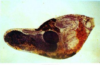
Diagnosis If the disease is endemic or the animal is from the endemic area of the disease, and in the onset season of the disease, the clinical manifestations of long-term wasting, anemia, repeated indigestion, the treatment effect is not obvious, that is, whether there is trematodiasis should be considered. To confirm the diagnosis, the eggs can be examined by stool washing and precipitation, and if necessary, the egg caterpillar hatching method can also be used. When the animal is posthumously autopsyed, if the worm is found in the hepatobiliary duct, the pancreatic duct, and the mesenteric vein blood vessel, the diagnosis can be confirmed.
Immunological diagnostic methods such as cyclic egg precipitation test, indirect agglutination test and enzyme-linked immunoassay have been applied in production practice.
Treatment The following drugs can be used according to the actual situation:
Praziquantel, cattle 35 to 45 mg per kilogram of body weight, sheep 60 to 70 mg per kilogram of body weight, once oral, or cattle, sheep are 30 to 50 mg per kilogram of body weight, with liquid paraffin or vegetable oil formulated into a sterilization oil, intraperitoneal injection.
HC-p-xylene (blood-846), cattle 300 mg per kilogram of body weight, sheep 400 to 600 mg per kilogram of body weight, oral, once every other day, 3 times for 1 course of treatment.
Trichlorobenzene *** (hepatic leech net), cattle per kilogram of body weight 10 mg, sheep per kilogram of body weight 12 mg, 1 oral, the drug for liver flukes adult and child worms are effective, the drug suspension period of 14 days.
Trichlorobenzoylazine (hydrochlorophenylpropionazine), cattle per kilogram of body weight 30 ~ 40mg, sheep per kilogram of body weight 40 ~ 50mg, formulated into a 2% suspension, oral administration, the drug has a special effect on the Qilu flu fluke.
Propiopyrimidium *** (anti-helminthiasis) is also commonly used in the treatment of liver flukes and chilis, with cattle 10 to 15 mg per kilogram of body weight and sheep 30 to 40 mg per kilogram of body weight.
Prevention In areas where the disease is endemic, pastures and grazing should be selected as much as possible in high-dry areas. It is best to perform two group-wide preventive dewormings in late autumn and early winter and early winter and early spring within one year. The elimination of intermediate hosts is an important part of the prevention of this disease, and can be carried out according to the biological characteristics of various intermediate hosts by chemical, physical, biological and other methods, but the impact on the environment should be fully considered. Sick animals and people should be dewormed and treated in time. Human and animal manure should be collected as much as possible and biothermal treatment to eliminate the eggs in it.