Late 19th century,
A farmer named Wilson Bentley was self-taught,
Attach the microscope on a folded camera camera,
The first microscopic photo of a single snowflake was successfully taken
And published in 1923 in National Geographic
——
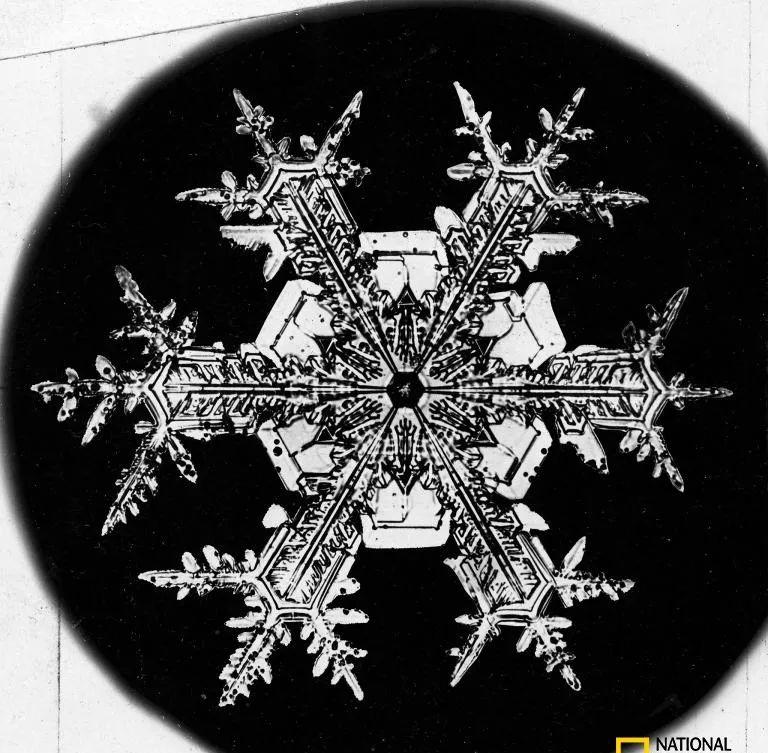
Photo by Wilson Bentley
from now on
The advanced realm of "a drop of water contains a world",
Each of us can see it with our own eyes.
Although the microscopic world is only a particle that ordinary people see under a microscope, it contains a huge cosmic system, which contains "suns", "mountains", "rivers", and life, as well as all the necessities of survival. On a grain of sand, there are thousands of beautiful villas.

To reproduce, the moss combines its sperm and egg to form a cell that develops into a stem with a sporangia attached to the tip (pictured as a cross-section of the cell). The spores mature in the sporangia, after which the sporangia burst open and the spores are emitted.
Photo by MAREK MIS, SCIENCE SOURCE
Until now,
Everything in the microscopic world is still amazing.
The object in the picture is not an oval earth pot filled with grass, but an immature sub-solid head of a dandelion flower under a scanning electron microscope.
摄影:THE NATURAL HISTORY MUSEUM,LONDON;SCIENCE SOURCE
In order to show everyone the most invisible miracles of art and science,
Nikon founded the Miniature World Micro Photo Contest in 1974.
And it goes on forever,
This year is the 46th edition.
The competition collected thousands of entries worldwide. Recently, a jury of scientists, photographers and journalists announced 20 award-winning works.
The picture shows the back of a live little zebrafish, which won the first prize in the 2020 Nikon Miniature World Micro Photojournalism Competition. To achieve surreal results, National Institutes of Health researcher Daniel Castranova, aided by colleague Bakry Samasa, labeled the fish's lymphatic vessels (orange) and scales (blue) with fluorescent proteins and stitched together more than 350 photos.
摄影:DANIEL CASTRANOVA, BRANT WEINSTEIN, BAKARY SAMASA, EUNICE KENNEDY SHRIVER NATIONAL INSTITUTE OF CHILD HEALTH AND HUMAN DEVELOPMENT,NATIONAL INSTITUTES OF HEALTH, NIKON SMALL WORLD
"We are very proud that these photographs combine research, creativity, imaging technology and expertise, and lead to new scientific discoveries," Nikon said. This year's championship is a shocking example. ”
The ethereal photograph above also leads to a groundbreaking scientific discovery: the presence of lymphatic vessels in the skulls of zebrafish is very similar to those of mammals and humans. This could help scientists better understand the human lymphatic system and make breakthroughs in neurological diseases such as Alzheimer's.
Most moths' eyes are covered with a layer of nanostructures that absorb light and prevent reflections, allowing them to better hide their movements. Taken by Indonesian photographer Ahmad Fauzan, this photograph shows a black optical sphere on the eyeball of an Asian-Australian white-skirted moth, along with its furry head and curly beak.
Photograph by AHMAD FAUZAN. SAIPEM, NIKON SMALL WORLD
German photographer Daniel Knop lined up five photos of clownfish embryos to show their development from hours after fertilization to hours before hatching.
摄影:DANIEL KNOP. NATUR UND TIER-VERLAG NTV, NIKON SMALL WORLD
The rainbow-shaped comb in the picture is actually the surface of the tongue (also known as the tooth tongue) of a freshwater snail, covered with tiny teeth that are used to grind food into a digestible size. Photo taken by Igor Siwanowicz of the Howard Hughes Medical Institute.
摄影:IGOR SIWANOWICZ. HOWARD HUGHES MEDICAL INSTITUTE, NIKON SMALL WORLD
Canadian scientists Vasileios Kokkoris, Franck Stefani and Nicolas Corradi photographed a soil fungus spore containing multiple nuclei. The spores are surrounded by a growing fungal fiber called hyphae.
摄影:VASILEIOS KOKKORIS, FRANCK STEFANI, NICOLAS CORRADI. UNIVERSITY OF OTTAWA & AGRICULTURE AND AGRIFOOD CANADA, NIKON SMALL WORLD
Robert Markus and Zsuzsa Markus of the University of Nottingham photographed large amounts of colourful pollen in the pollen sac of a long-flowered plant.
摄影:ROBERT MARKUS, ZSUZSA MARKUS. UNIVERSITY OF NOTTINGHAM, NIKON SMALL WORLD
Jason Kirk of Baylor School of Medicine photographed brightly colored nuclei (blue-green) surrounded by microtubules (orange) in a human cell.
摄影:JASON KIRK. BAYLOR COLLEGE OF MEDICINE, NIKON SMALL WORLD
Allan Carrillo-Baltodano and David Salamanca of Queen Mary University of London recapitulated autofluorescent chameleon embryos in black and white images. Fully developed chameleons can change their color and pattern through reflective crystals in skin cells.
摄影:ALLAN CARRILLO-BALTODANO, DAVID SALAMANCA. QUEEN MARY UNIVERSITY OF LONDON, NIKON SMALL WORLD
The hippocampus of the human brain is primarily responsible for memory and learning. This image, taken by Jason Kirk and Quynh Nguyen of Baylor Medical School, shows two adjacent hippocampal neurons connected together by elongated synapses, ready to complete some memory activity.
摄影:JASON KIRK, QUYNH NGUYEN. BAYLOR COLLEGE OF MEDICINE, NIKON SMALL WORLD
Macrocampus is a small crustacean with a transparent shell that reproduces asexually, so it is often used as an experimental organism. Pictured here is Ahmad Fauzan's second award-winning work, showing the head of one of the large clumps.
The Red Algae tendrils photographed by tagide de Carvalho at the University of Maryland stretch out like skull fingers, as if to grasp something.
摄影:TAGIDE DECARVALHO. UNIVERSITY OF MARYLAND, ALTIMORE COUNTY, NIKON SMALL WORLD
Austrian photographer Robert Viethaler superimposes many pictures together to show a strand of knotted hair.
Photograph by ROBERT VIERTHALER. NIKON SMALL WORLD
In this work by New York photographer Justin Zoll, the crystals formed by the heating of ethanol, water and amino acid solutions are delicately intertwined to form a beautiful flower pattern that looks like an abstract painting by Georgia Okeeffer.
摄影:JUSTIN ZOLL. JUSTIN ZOLL PHOTOGRAPHY, NIKON SMALL WORLD
In this work, taken by Turkish photographer Ozgur Kerem Bulur, a leaf-curling weevil climbs up a plant under the protection of a textured, reflective shell.
Photograph by ÖZGÜR KEREM BULUR. NIKON SMALL WORLD
In this work, taken by Argentine scientists Eduardo Zattara and Alexa Bely, an asexually reproduced clear-cut caterpillar produces a cascade of offspring.
摄影:EDUARDO ZATTARA, ALEXA BELY. NAC INSTITUTE. RESEARCH ON BIODIVERSITY AND ENVIRONMENT, NIKON SMALL WORLD
This interlocking, tangled color mesh photograph, delicately reimagined by Russian photographer Alexander Klepnev, is actually a very ordinary thing: a nylon stocking. This is the only non-natural object photography work that has won an award in this year's photo contest.
摄影:ALEXANDER KLEPNEV. JSC RADIOPHYSICS, NIKON SMALL WORLD
Britain's Anne Algar superimposed many pictures to create this stitching of cold glowing scratches. The scratch bug is an aquatic insect that spreads around the globe.
Photograph by ANNE ALGAR. NIKON SMALL WORLD
In this work by California photographer Chris Perani, the wings of an Atlas moth are covered with a layer of soft scales that look like thousands of small feathers.
Photo by CHRIS PERANI. NIKON SMALL WORLD
Jan Michels of the University of Kiel in Germany photographs a photograph of a marine diatom with a patterned cell wall. Diatoms produce more than 20% of the earth's oxygen each year.
摄影:JAN MICHELS. CHRISTIAN-ALBRECHTS-UNIVERSITÄT ZU KIEL, NIKON SMALL WORLD
Dorit Hockman and Vanessa Chong-Morrison of the University of Cape Town took photographs of the brightly colored skeleton of the short-tailed lobed epistaxis embryo.
摄影:DORIT HOCKMAN, VANESSA CHONG-MORRISON. UNIVERSITY OF CAPE TOWN, NIKON SMALL WORLD
If you haven't seen enough, let's take a look back at the wonderful photography of the 45th edition -
Microscopy technologist Teresa Godard and student Teresa Coogler won the championship by stitching together hundreds of images to finally synthesize this fluorescent turtle embryo image. Embryos are stained to distinguish between different tissues.
Photography: TERESA ZGODA, TERESA KUGLER
Iger Svanovicz, a scientist at the Howard Hughes Institute of Medicine, applied color-coded imaging to the trumpetworms, a single-celled freshwater protozoa, to highlight the 3D shape of these organisms.
摄影:IGORSIWANOWICZ, HOWARD HUGHES MEDICAL INSTITUTE (HHMI) JANELIA RESEARCH CAMPUS
Jan Rosenboom of the University of Rostock in Germany combined several photographs taken at different focal lengths to synthesize a picture of the male mosquito.
In the microscopic world, all the details are magnified, things seem to have changed their appearance, who would have thought that such a "fairy fluttering" look would be the most hated mosquito.
Purchase the 2021 National Geographic Chinese calendar