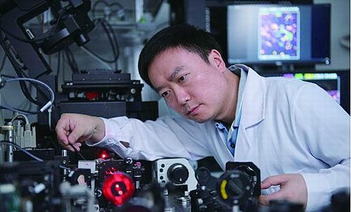
Li Dong at work (Photo courtesy of interviewee)
Green is the mitochondria, purple is lysosomal, and yellow is the microtubules.
The mitochondria move briskly along the linear microtubules as if gliding on their own light rails. If it weren't for the fluorescent markers that vividly stain each structure, it's easy to overlook the tiny lysosomes that connect them. These humble little guys are the "big guys" who recruit microtubules for their own use, and the big mitochondria are just a free ride.
It's a scene in a mammalian cell. The cell is attached to a small transparent box wall and placed under a skimmed incident structured light illumination microscope (GI-SIM).
Li Dong, a researcher at the Institute of Biophysics of the Chinese Academy of Sciences, sat in a dark laboratory, looking through a computer screen at the bustling scenes of this tiny world. He knew that this scene had never been seen before.
Look, life!
Over the years, Li Dong has led the team to develop multimodal structured light super-resolution microscopy, deep learning super-resolution imaging algorithms and three-dimensional high-space-time resolution biodynamic microscopy, and through cooperation with other biologists, they continue to show the wonders in the hidden corners of life.
Li Dong studied optical engineering at Zhejiang University as an undergraduate, and the first lesson of admission was to enjoy the scene of China's first atomic bomb explosion captured by the predecessors of the department with their own high-speed cameras.
Optical research is a "seeing" science, which is committed to expanding the limits of human vision: seeing what was once invisible, seeing what was once invisible, seeing what was not clear in the past, retaining fleeting images, putting eyes on lifeless machines...
An important lifelong choice for optical researchers is: What do I want to help the world see?
When Li Dong was studying for his Ph.D. at the Hong Kong University of Science and Technology, he was exposed to the interdisciplinary discipline of "biophotonics". After graduating with his Ph.D., he went to the United States to do postdoctoral work at the prestigious Howard Hughes Institute of Medicine in the life sciences under the supervision of Eric Betzig, who later won the Nobel Prize in Chemistry for his super-resolution fluorescence microscopy.
In the Betzig laboratory, Li Dong made the first major scientific breakthrough: successfully developed a high-value aperture nonlinear structure light illumination microscope, which increased the optical resolution of high-speed imaging of live cells to 60 nanometers. The result appeared on the cover of Science magazine in 2015.
This technology gives scientists the opportunity to clearly see the fine dynamics of life activity in living cells. Nature-Methodology comments: "This is an important step towards the eventual realization of observing life processes at molecular-level resolution!" ”
Among the alumni who graduated from the Hong Kong University of Science and Technology, Li Dong's choice is very small: "Many of my friends have done drones, and many people have not even heard of the Howard Hughes Medical Institute." But he was convinced of this choice: "I gradually realized that the life sciences are a meeting point for various disciplines, a big stage with infinite possibilities." ”
Dance with "Restrictions"
When he first started doing biological optical imaging research, Li Dong was often confused by a problem: what he observed was not what biologists needed most. Many times, it's not that the clearer it is, the more valuable it is.
The cell structure is three-dimensional, but to observe the dynamic changes of life, it also needs to be extended to the "five dimensions" - in addition to x, y, z (spatial dimensions), adding time dimensions and color dimensions. Without a color dimension, organelles such as mitochondria, lysosomals, and microtubules cannot be distinctly distinguished; without a time dimension, they cannot capture their rapidly changing activity.
"But the relationship between these five dimensions is 'mutually exclusive.'" Li Dong said, "It is impossible for the index performance of each dimension to be high, and while ensuring the time resolution and color resolution, it is necessary to sacrifice a part of the spatial resolution." ”
It's an art of balance and compromise — to observe colorful dynamics, you have to give way to some imaging speed. How to obtain the best comprehensive effect at the smallest possible cost is the goal that Li Dong's team has always pursued.
Objective law limitations like this are ubiquitous in research.
The limitation of electron microscopy is that it is impossible to observe dynamic living objects. When Li Dong turned his gaze to the optical microscope, he encountered the "Abbe Limit" that was difficult to break through.
The limit of optical microscopy resolution is about half the wavelength of visible light. Calculated by the shortest wavelength of blue-violet light in visible light, it is half of the wavelength of 400 nanometers - 200 nanometers. This resolution is far from enough to see biological macromolecules of a few nanometers and tens of nanometers.
Eventually, Li Dong found inspiration from the formula he learned in high school: two sine functions are multiplied, and the frequency of the wave function increases. In practice, it is to give two different colors of light sources, so that their peaks and peaks are superimposed on each other, so as to break through the original limit and greatly improve the resolution.
"The Abbe limit still exists, and existing technical methods can only bypass it." Li Dong smiled. In the face of the laws of nature and the flaws of reality, "circling" is an indispensable wisdom. In another study, Li Dong correlated images obtained under low signal-to-noise ratio shooting conditions with images with high signal-to-noise ratios, and by letting machine learning this mapping, it is possible to convert relatively ideal results from undesirable images.
Between two points, sometimes the curve is the shortest.
It's the ultimate and the beginning
Li Dong said that the principles he used were not difficult to imagine. The real difficulty lies in how to do it perfectly.
"For 11 years, we have developed and polished this microscope system that integrates so many illumination modules together, each of which can achieve optimal specifications without compromising the imaging performance of other modules, which may be unique in the world." Li Dong, who has always been restrained in talking, said this, and for the first time, he showed a little pride.
In Li Dong's laboratory, you will see a "microscope" that is completely different from what you imagined. It is not a complete, smooth and decent finished product, but a complex link composed of a large number of optical components that interlock and rotate thousands of turns. It's like spreading out "internal organs", which at first glance makes people dizzy.
The position, angle, height and height of each component have been carefully debugged, and the whole body is involved. You can't be wrong, once you lose a millimeter, it will be a thousand miles.
"Every optical component is not perfect, it has a certain so-called 'aberration'. The trick is not to optimize the accuracy of each link through complex design, but to cancel out and compensate each other for the errors between the links. Li Dong said.
The incomparably complex and exquisite microscopic imaging system created by ten years of grinding a sword has welcomed collaborators and graduate students from various institutions and different professional backgrounds, and has welcomed a wave of curious and ambitious explorers.
But Li Dong is very sober, he knows that these achievements only provide potential technical storage and support for the "short board" and "stuck neck" problems in the field of high-end optical microscopy imaging equipment in the mainland. "All our work has yet to be tested in biological research and verified by the laws of the market. Our real value is ultimately reflected in helping biologists achieve cutting-edge scientific results. ”
From the beginning of the use of fluorescence, to the first fusion of fluorescent proteins into the proteins of living organisms, and then to the wide application of fluorescence in life science research and microscopy technology, it took 80 or 90 years.
The road of scientific research and innovation is so long and long. Taking climbing Mount Everest as an analogy, Li Dong believes that he has just set off, "may not have reached the base camp, and maybe even where the base camp is to be explored."
"But what is different from scientific research is that we do not have a mature route that has been formed, only through our own exploration, to explore every rock, every ditch, to pursue more ideal microscopic imaging tools, to discover more amazing life secrets." He said.
(Reporter Li Chenyang)
Source: China Science Daily
![Do things with force, vigorously produce miracles, such as seeing a person at a great distance, making an effort to get close to her, as long as you have gained all the strength of it, and trying to get closer to her, you are already paying, such as[fig]](data:image/gif;base64,R0lGODlhAQABAIAAAP///wAAACwAAAAAAQABAAACAkQBADs=)