This article is from the WeChat public account: X-MOLNews
"Raising single crystals" is a topic that makes many small partners in the field of chemical materials sad. How many people, the paper data is inferior to a single crystal structure, but is depreciated by the picky reviewers? How many people, raising single crystal sieve conditions to screen the amount of hair sharply reduced, but by the hands of fine, broken, miscellaneous microcrystals to doubt life?
The root cause of all this lies in the current "gold standard" for structural analysis – single crystal X-ray diffraction (XRD) – of high demands on samples: single crystals that are large in size, have few defects, are sufficiently stable and are not sensitive to radiation. This is an almost impossible task for some compounds. For example, inorganic organic hybrid materials that have been popular in recent years can often only get fine microcrystals, low symmetry and sensitivity to radiation, and can not be on XRD at all.
Recently, the University of Connecticut J. Nathan Hohman and Nicholas K. Sauter and Aaron S. Brewster at Lawrence Berkeley National Laboratory report a new approach, small-molecule serial femtosecond X-ray crystallography (smSFX), combined with high-throughput, Femtosecond pulsed X-ray free electron lasers (XFEL) and novel data processing algorithms enable the acquisition of high-resolution structures of microcrystalline metal-organic materials. This method does not require the material to be resistant to radiation, requires little to no sample preparation process, and can be performed at room temperature and atmospheric pressure, theoretically suitable for structural resolution of all microcrystallines. The paper was published in the journal Nature.
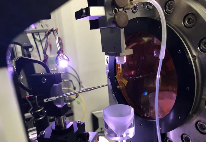
Part of the XFEL installation. Image credit: University of Connecticut [1]
Powder diffraction technology is often used for microcrystalline structure studies, but because the sample is actually mixed together countless microcrystallines, the mixture diffracts together, can only get the mixed electron density data, it is difficult to perform high-resolution analysis. The serial femtosecond crystallography (SFX) method is different, with the help of XFEL's high flux and femtosecond pulses, it can achieve "diffract before destruction", obtaining diffraction data in a single frame of a single submicron crystal, the so-called "single shot per crystal". To take a less appropriate example, powder diffraction is like a cannon fire covering shooting, hitting a large group at a time, but the effect is difficult to evaluate; SFX is like a precision weapon for fixed-point strikes, one at a time, one at a time, one data at a time, and one by one. Overall, SFX has no small advantages, for example, the data collection duration of a single microcrystalline is only on the femtosecond scale, avoiding the destruction of microcrystallines by radiation; data collection can be carried out under room temperature pressure, without freezing or vacuum; and data collection of the entire sample takes only a few minutes, saving time and money.

This paper smSFX method and the morphology of the three research objects. Image credit: Nature
However, SFX's previous use in the chemical field of crystal structure analysis was not without its challenges, and the problems often focused on unit cell assays and diffraction pattern calibration. In a traditional single XRD process, the crystals rotate on a goniometer and the crystal orientation matrix is known, while in the SFX process, each crystal in the suspension is randomly oriented, so the orientation matrix must be determined separately for each frame (Figure c below). In macromolecular SFX, macrocrystal cells produce dozens of reflections per frame, with significant periodicity, which can be calibrated using the Fourier method. Unlike small molecule SFX, which has only 3-10 reflections per frame, this sparsity requires the use of calibration algorithms that do not rely on periodicity. The authors' team has made important progress on this issue, with the help of advanced algorithms (relevant code and descriptions have been made public)[2], synthesizing high-resolution powder diffraction patterns from XFEL data to determine unit cells, calibrating sparse diffraction patterns by graph theory methods to generate data sets, and then using standard tools for single crystal diffraction data to analyze and refine the high-resolution crystal structure of microcrystalline samples.
Powder diffraction pattern synthesized from XFEL data. Image credit: Nature
The samples studied in this paper are three microcrystalline metal-organic hybrid sulfur compound materials mithhrene (AgSePh), thiorene (AgSPh) and tethrene (AgTePh). Based on the smSFX method, the structures of all three materials were successfully obtained. The Mithrene structure has a resolution of 1.2 Å and is consistent with the previously reported single crystal structure.
Mithrene structure determination results and previously reported single crystal structure (d). Image credit: Nature
As for Thiorene and tethrene, which have never reported a crystal structure before, a high-resolution structure of 1.35 Å was also obtained based on the smSFX method. Further analysis showed that the different optoelectronic properties of the other two materials exhibited by the thiorene material were related to their different Ag-Ag bonding networks.
Crystal structure of three materials and Ag-Ag network. Image credit: Nature
The above results show that smSFX is expected to become a general technique for determining the structure of radiation-sensitive microcrystalline materials at temperatures and pressures close to the environment. Advances in crystallography are also expected to help scientists design better materials from scratch.
PS: Turn over the experiment of not being able to raise a single crystal before turning over, and see if you can still save it with smSFX?
Chemical crystallography by serial femtosecond X-ray diffraction
Elyse A. Schriber, Daniel W. Paley, Robert Bolotovsky, Daniel J. Rosenberg, Raymond G. Sierra, Andrew Aquila, Derek Mendez, Frédéric Poitevin, Johannes P. Blaschke, Asmit Bhowmick, Ryan P. Kelly, Mark Hunter, Brandon Hayes, Derek C. Popple, Matthew Yeung, Carina Pareja-Rivera, Stella Lisova, Kensuke Tono, Michihiro Sugahara, Shigeki Owada, Tevye Kuykendall, Kaiyuan Yao, P. James Schuck, Diego Solis-Ibarra, Nicholas K. Sauter, Aaron S. Brewster & J. Nathan Hohman Show fewer authors
Nature, 2022, 601, 360–365, DOI: 10.1038/s41586-021-04218-3
Resources:
1. New X-Ray Technique Sees the Crystal in the Powder
https://today.uconn.edu/2022/01/new-x-ray-technique-sees-the-crystal-in-the-powder/
2. Code and instructions for diffraction data processing
https://github.com/cctbx/cctbx_project/tree/master/xfel/small_cell