<h1>summary</h1>
Animal parasites are tiny insect bodies that parasitize inside and outside animals, infect and seize nutrients through pathogens, and endanger humans and animals.
<h1>First, parasite cognition</h1>
1. Single-host parasite: only one host is needed, such as: roundworms, hookworms; that is, soil-derived parasites.
2. Multi-host parasites: multiple hosts are required, such as: polyester worms, trematodes; that is, biogenic parasites.
3. End-host: refers to the animal parasitic by the insect body in the stage of sexual maturity or sexual reproduction.
4. Intermediate host: refers to the animal body parasitic in the larval stage or the asexual reproductive stage; the pig is the intermediate host of the pig with polyester worm.
5. Supplementary host: that is, the second intermediate host; for example, the supplementary host of biluminal flukes is ants.
6, storage host: that is, the transport host, refers to the parasite in the host body does not develop and reproduce but has infectious power, this host is called "storage host". For example, the storage host of chicken heterogeneous nematode is earthworm.
7. Carnivote host: refers to the host that is not accustomed to being parasitic; for example, the host of the carnivost of schistosomiasis is a cultivating cow.
8. Worm-bearing host: refers to the host that is in a state of hidden infection after being infected, has no clinical symptoms, and has a certain immunity to re-infection.
9. Vector: refers to a type of animal that transmits parasitic diseases among vertebrate hosts; such as mosquitoes and insects.

10. Pathogenesis of parasitic diseases:
(1) Plundering nutrients, such as: roundworms, polyester insects.
(2) Mechanical damage; such as: roundworms.
(3) Insect toxins and immune damage, such as: Chinese mycodystis, schistosomiasis.
(4) Secondary infection, such as: flea-transmitted Y. pestis.
11. Direct developmental type: refers to the developmental history that does not require an intermediate host.
12. Indirect developmental type: refers to the developmental history of the need for intermediate hosts.
13. Route of infection: oral, skin contact, arthropod, transplacental, self-infection.
14. One of the main characteristics of parasitic diseases: chronic infection.
15. Hidden infection: refers to the infection of parasites, no clinical manifestations, conventional methods can not detect.
16. Potential period: the shortest time from the time the parasite infects the host to the time the next generation of eggs are discharged.
17. Immune evasion mechanism of parasites:
(1) Histological isolation, such as: Toxoplasma gondii in the fetus; intracellular parasites.
(2) Changes in surface antigens, such as: split flukes.
(3) Immunosuppression, such as: trypanosomiasis, liver bovine flukes.
(4) The production of soluble antigens, such as: visceral Lechermania.
(5) Metabolic inhibition, such as: intracellular toxoplasma, Trypanosoma subtilis.
Second, the diagnosis and prevention and control technology of parasitic diseases
1. The most reliable diagnostic method for parasitic diseases: pathogen examination.
2. Commonly used fecal testing methods:
(1) Naked eye observation.
(2) Direct smear method.
(3) Egg floating method; (used to check nematode eggs, polyester eggs and coccidiosis oocysts to establish a prenatal diagnosis).
(4) Egg precipitation method.
(5) Egg counting method (commonly used McMaster's method).
(6) Larval culture method.
(7) Larval separation method.
(8) Caterpillar larval hatching method.
3. Nematode eggs: The egg shell is composed of two layers, and there are egg cells in the shell, and the egg shell of the roundworm egg is the thickest.
4. Trematodes eggs: the egg is round, and the egg shell is composed of several layers of membranes, thick and solid.
5, polyester worm eggs: the center of the worm eggs have an oval shape with three pairs of embryo hooks of the six hooks (embryos), mostly colorless.
6. Diagnosis of scabies mites: at the junction of the affected part and the health department, the epidermis is repeatedly scraped with a convex blade knife, and the scraped dander is added to the 50% glycerol solution for post-microscopic examination.
7. Examination of demodex mites: use a knife to cut the sandy nodules on the skin, pick the purulent secretions on the carrier, add normal saline, and then microscopically examine.
8. Examination of lice, ticks and fleas: the parasites of animal armpits, rat moles, breasts and toes and behind the ears are collected with forceps and examined by post-mirror with transparent treatment.
9. Blood parasites: including trypanosomiasis, babesia, Taylor's worm, resident leukoplasma and heartworm.
10. Examination method of blood parasites: (1) Blood smear is stained. (2) Observation of blood pressure drops (check the movement type of the insect body). (3) Insect body concentration method (used to check Trypanosoma eliii and P. piriformis).
11. Examination of bovine fetal trichomoniasis: (1) The disease materials that can be collected are: transparent mucus secreted by the vagina, bull foreskin irrigation fluid, the fourth gastric contents of the miscarried fetus or chest and ascites; (2) the undissigned specimen microscopy can clearly see the fluctuating membrane.
12. Examination of equine trypanosomiasis: the disease materials that can be collected are: (1) the skin or papule extraction of the puffy part, the scraping of the mucous membrane of the urethra and vagina; (2) the mucosal scraping is the most likely to find the insect body.
13. Examination of Taylor's worm: lymph node puncture smear, fixation, and staining microscopy to detect Koch's blue body.
14. Examination of toxoplastic bodies: (1) microscopic examination of sacs or tachygospora after death of sick animals; (2) diagnosis before death can be taken for ascites, smears, and microscopic examination of trophozoites.
15. Examination of trichinella: (1) Microscopic examination: cutting the diaphragm into small pieces for posterior microscopic examination. (2) Digestion method: take 100g of meat sample, add 10g of pepsin, dissolve in 2L tap water, and add 16mL of hydrochloric acid (25%).
16. The selection principle of anthelmintic drugs: high efficiency, low toxicity, broad spectrum, inexpensive and easy to use.
17. Deworming time: Generally "before the insect body matures", or "autumn and winter". The feces excreted after deworming are treated with "biothermal fermentation".
18. Formula for evaluating the efficacy of anthelmintic drugs:
(1) The rate of egg to negative = the number of animals with eggs turning negative / the number of experimental animals multiplied by 100%.
(2) Egg reduction rate = (pre-deworming EPG - post-deworming EPG) divided by pre-deworming EPG multiplied by 100%.
(where EGP: the number of eggs per gram of feces)
(3) Sperm deworming rate = number of excreted insect bodies divided by (number of excreted insects + number of residual insect bodies) multiplied by 100%
(4) Rough deworming rate = (average number of residual insect bodies in the control group - average number of residual insect bodies in the experimental group) / The average number of residual insect bodies in the control group multiplied by 100%.
(5) Expulsion rate = exorcism of insectoid animals divided by the total number of test animals multiplied by 100%
19. Deworming of poultry: first mix the drug in a small amount of wet material, then mix it into the daily feed, and feed it after 6-12 hours of hunger strike.
3. Zoonotic parasitic diseases
1. Toxoplasmosis: (1) The worm body parasitizes the nucleated cells of the host, with only one clock and one serotype.
(2) Includes 3 stages of development: trophoblasts, cysts, and oocysts.
(3) Nourishing type: banana-shaped, blue cytoplasm after staining, purple nucleus, mostly found in blood, cerebrospinal fluid, pathological exudate in the acute stage.
(4) Cystic, oval, containing multiple bradys, most common in the brain, skeletal muscle, myocardium in chronic cases.
(5) Egg sac: round, mostly found in the feces of the terminal host cat.
(6) Symptoms: pig hyperthermia, constipation or diarrhea, vomiting, dyspnea, body surface lymphadenopathy, cyanosis of the ears and abdomen, stillbirth or miscarriage in pregnant pigs.
(7) Diagnosis: Take the blood during the fever period and detect it with immunofluorescence.
(8) A positive specific IgM represents early infection.
(9) Treatment: sulfonamides.
(10) Prevention: The disease is caused by the ingestion of egg sacs in cat feces; that is, it is strictly forbidden to raise cats and prevent cat feces from contaminating feed and drinking water.
(11) Acute lesions: lungs, hepatic lymph nodes, kidney swelling and hard necrosis, systemic bleeding, etc.
2. Leishmaniasis:
(1) That is, black fever, death can reach 40%; China has now controlled the disease.
(2) It is parasitic in the endothelial cells of dogs and is transmitted by whiteflies, and its natural host is dogs.
(3) Symptoms: skin type (the superficial layers of lips and eyes can heal by themselves), visceral type (hair removal eczema, protozoa are abundantly present in the skin).
(4) Pathology: swelling of the spleen and lymph nodes.
(5) Treatment: antimony preparation.
3. Japanese schistosomiasis:
(1) That is, "schistosomiasis", humans, cattle, sheep, pigs as the main source of infection.
(2) Epidemic areas: Yangtze River Delta (water network type), middle and lower reaches of the Yangtze River (the highest rate of limnological infection), except for Shanghai overseas provinces and regions (hill type).
(3) Adult parasitism occurs in the portal vein and mesenteric vein, and the tail rush is mainly infected by the skin of the fetus.
(4) Spring and summer are frequent, and the infection rate is the highest among young adults.
(5) Symptoms: elevated body temperature, irregular intermittent fever, diarrhea or blood in the stool, wasting, developmental delay, anemia, exhaustion and death, cow miscarriage, calves become pygmy cattle.
(6) Lesions: more common in the intestine, liver, spleen, the basic lesions are worm egg nodules caused by egg sedimentation in the tissue.
(7) Diagnosis: fecal caterpillar hatching method, indirect hemagglutination test.
(8) Insect body characteristics: hermaphroditic, parasitized male and female hugging, the insect body is long cylindrical, the appearance of linear.
(9) Treatment: praziquantel; snail control: chloroniclosamide ethanolamine salt.
(10) Prevention: prevention first, prevention and control combined, classified management, comprehensive management, joint prevention and control, human and animal co-treatment, and key management of infectious sources.
4. Cysticercosis:
(1) It is a larva of pig-belt polyester worms in human body, and is one of the key projects of meat hygiene inspection.
(2) Form: The larvae of the pig cystic tail larvae are oval;the adult worms are pig band polyester worms, or "hooked polyester worms"; the eggs are round and contain six hooked worms.
(3) Intermediate host: pig, terminal host: human.
(4) Symptoms: Generally there are no obvious symptoms, and pigs are mostly caused by eating feed contaminated by humans.
(5) Diagnosis: Among the muscles, the detection rate is the highest in the muscle groups on the lateral side of the anterior arm.
(6) Prevention: Grasp the five links of "investigation, driving, inspection, management, and reform".
(7) Treatment: praziquantel, imidazole propylthiazole.
5. Echinococcosis:
(1) That is, "echinococcosis", is a polyester stage larvae of the echinococcal (cymbal) polyester worm in the small intestine of the terminal host dog, and sheep are the most susceptible and mostly parasitize in the liver and lungs.
(2) Pathogenic effect: mechanical compression, toxin effect, allergic effect.
(3) Prevention: It is forbidden to feed dogs with sick and dead liver and lungs.
(4) Treatment: imidazole propylthiazole, pyroquine.
(5) Confirmed diagnosis: Autopsy is found in the liver and lungs.
6. Trichinella:
(1) One of the meat health quarantine items: adults parasitize the small intestine, and larvae parasitize the striated muscle.
(2) The main source of infection: pigs, dogs, cats, rats are the main source of infection of swine trichinellosis, pigs are the main source of infection of human trichinellosis, and human infection with trichinella is mostly caused by eating raw or eating undercooked meat.
(3) Diagnosis: tablet microscopy (cut 24 grains of meat sample the size of wheat grains, microscopic examination of the larvae of the capsule, the capsule is fusiform, and there are curled insect bodies in the capsule).
(4) Muscle digestion method.
(5) Treatment: imidazole propithiazole.
Fourth, a variety of animals co-exist with parasitic diseases
1. Trypanosomiasis:
(1) "Sula disease", parasitic in the blood, lymph nodes, and hematopoietic organs of horses and cattle; propagation by longitudinal splitting; transmitted by insect death.
(2) Horses and dogs are the most susceptible, with frequent occurrences from July to September.
(3) Clinical features: subcutaneous edema on the chest, crotch and penis.
(4) Horse: acute passage, high death, body temperature of more than 40 degrees, after a few days of normal return, and then rise, so repeated; wasting, anemia, jaundice, transient membrane seeing hemorrhagic spots, dedema of the body hypoptosis.
(5) Beef liver then shows chronic passage.
(6) Diagnosis: The detection of insects in the blood is the most reliable diagnostic basis; such as: pressure drop specimen examination.
(7) Prevention and control: benzenesulfonylurea, quinopyramide, nitrogen chloride, phenidine hydrochloride.
(8) Clinical use of drugs for prevention.
2. Neosporidiosis:
(1) Oocyst: in the intestine of the terminal host dog, reticulated.
(2) Tachygotes (trophozoites) are present in the placenta of the intermediate host cattle.
(3) Cyst: it exists in the nervous system of newborn calves and is rounded.
(4) Symptoms: cattle abortion, calf congenital neuromuscular injury.
(5) Lesions (pathology): non-purulent encephalomyelitis.
(6) Diagnosis: specific antibody staining, indirect immunofluorescence test.
(7) Prevention and control: eliminate sick cattle and seroant antibody-positive cattle.
3. Cryptosporidiosis:
(1) Symptoms: Severe diarrhea in calves and lambs, severe respiratory symptoms in poultry, and severe diarrhea in humans, which are of great public health significance.
(2) Development: experiencing oocysts, sporozoites, fissocids, trophozoites, gametophytes, gametes, etc.
(3) Water pollution is an important cause, and it occurs frequently in wet and warm seasons.
(4) The most common is Cryptosporidium annii in dairy cows, and the most widely used in poultry.
(5) Diagnosis: Oocysts in feces are collected by saturated sucrose liquid floating method, and rose-red oocysts are detected under oil microscopy.
(6) Take the mucosa of the digestive tract of the dead case. Make a smear, stain it with Zinel's staining, and observe the round red insect body on a green background.
(7) Prevention and control: the effective treatment of feces and environmental health control are the most effective,
(8) Disinfectant: ammonia or formalin.
(9) The oocyst contains 4 bare sporozoites or 1 large remnant.
4. Mycosporidium disease:
(1) Terminal host: canine (small intestine epithelial cells).
(2) Intermediate host: herbivores (intramuscular).
(3) Diagnosis: the yellow-white cyst in the muscle can be seen with the naked eye, and the banana-shaped slow breeder or fecal examination of the oocyst or sporangia is examined by the posterior microscopy of the cyst.
(4) Prevention: Dogs and cats are strictly prohibited from approaching livestock and poultry.
5. Chinese mycodyloidosis:
(1) Parasitism in the liver, gallbladder, and bile ducts of terminal hosts, dogs, cats, and pigs.
(2) The insect body is flat and leafy, and the adult life span can reach 20 years.
(3) The first intermediate host --- freshwater snails, the second intermediate host --- freshwater fish and shrimp, and people, pigs, dogs, and cats are infected by swallowing such fish and shrimp.
(4) Symptoms: Mostly latent infections.
(5) Diagnosis: "Centrifugal fecal egg detection" has the highest detection rate, (the upper end of the egg has an egg cap, and the lower end has a small protrusion, containing worms)
(6) Treatment: praziquantel, propioimidazole, hexachloro-p-xylene.
(7) Prevention: Military dogs should have regular fecal inspections and not eat raw fish and shrimp.
6. Roundworm disease:
(1) That is, "rodworm disease", the first stage larvae are rod worm type, and the second stage larvae are filarial worm type (infectious to animals).
(2) The insect body in the animal is a female insect, and the insect body of the free generation lives in the soil, and the eggs are round and thin, containing knife-like larvae.
(3) His life history is alternating generations.
(4) Main route of infection: skin.
(5) Symptoms: 1-month-old piglets are the most susceptible, emaciated, growth retardation, and mass death.
(6) Orchidoid protodes parasitize the small intestine of pigs, especially in the mucosa of the duodenum.
(7) Pathology: eczema, alveolar bleeding, catarrhal or edema or ulcerative enteritis.
(8) Diagnosis: Fecal examination of worm eggs can confirm the diagnosis.
(9) Treatment: thiobenzozole.
7. Hairy tail nematode disease:
(1) That is, "hairy nematode disease" and "whipworm disease", the insect body is milky white, the male is curved at the back, the female is blunt and rounded at the rear end, the eggs are brownish yellow, the waist is drum-shaped, the egg shell is thick, and there are plugs at both ends.
(2) Mainly parasitic in the cecum, the highest infection rate at the age of four months, mostly summer infection, autumn and winter symptoms.
(3) Lesions: blind, colonic extensive chronic catarrhal inflammation.
(4) Diagnosis: fecal examination of worm eggs or autopsy to find worm bodies.
(5) Treatment: left imidazimid, benzthiazole.
8. Scabies mite disease:
(1) Chronic skin diseases parasitized by scabies mites in the epidermis, adult worms are round, have 4 pairs of limbs, adult worms dig tunnels in the host skin, and lay eggs and hatch into larvae, and the larvae are transformed into nymphs and adults.
(2) Features: severe itching, eczematous dermatitis, hair removal, the affected part expands to the periphery, highly contagious, mostly occurs in the head.
(3) Diagnosis: Collect disease materials at the junction of diseased skin and healthy skin, and microscopically examine insect bodies.
(4) Treatment: ivermectin, avermectin, cypermethrin, diazinon.
(5) Scabies mites, severe itching, hair removal, scabs, cracking.
9. Itch mite disease:
(1) Caused by mite parasitism on the surface of the skin.
(2) Eggs, larvae, nymphs and adults are all completed on the surface of the livestock body, and the body fluids are sucked as nutrients.
(3) Sheep, cattle, rabbits, the most susceptible, of which the harm to sheep is particularly serious, autumn occurs frequently.
(4) Symptoms: strange itching, goats mainly occur on the inside of the ear shell, buffalo body surface formed "paint exposure" like scab, rabbit otitis externa.
(5) Scraping the dander microscopy to examine the worm body.
(6) Treatment: avermectin.
10. Tick disease:
(1) It is an important blood-sucking parasite that parasitizes the surface of livestock and poultry.
(2) Hard tick: reddish brown, with a shield plate on the back, parasitizes in small mammals at the larval and nymph stages, and adults parasitize in livestock, mostly in the warm season.
(3) Soft tick: there is no shield on the back of the body, it is elastic leather-like skin, its life history is incompletely perverted, through the four stages of eggs, juveniles, if and adults, living in the gaps of livestock and poultry houses, mostly sucking blood in the middle of the night.
(4) Diagnosis: Ticks are found in animals to confirm the diagnosis.
(5) Prevention and control: ivermectin, cypermethrin, diazine farmers.
Fifth, the parasitic disease of pigs
1. Coccidiosis:
(1) A protozoal disease caused by Emmy coccidiosis and isosporangia parasitize pig intestinal epithelial cells.
(2) 7-21 day old piglets have multiple benign processes.
(3) Development: in the host epithelium, intracellular fission reproduction and gamete reproduction stages, spore reproduction in the outside world, the disease is transmitted through the digestive tract.
(4) Symptoms: piglet yellow-white, paste-like diarrhea, the most serious in July-August.
(5) Pathology: yellow fibrous and necrotizing pseudomembrane appear in the mucosa of the jejunum and ileum.
(6) Diagnosis: 7-14 days to make pig diarrhea with antibiotics ineffective, saturated saline floating method fecal examination of oocysts.
(7) Prevention: Doing a good job in environmental hygiene is the best way.
(8) Treatment: 100 balls clear.
2. Pork ginger flakes trematodiasis:
(1) Parasitizes pigs and human duodenum, causing diseases that affect the growth and development of piglets and the health of children.
(2) Form: Fresh insect body is fleshy red, and the insect body is thick and oblong,ovoid, like a thick slice of ginger cut obliquely.
(3) Intermediate host: flat snail (pond is its optimal growth environment, pigs are infected by eating aquatic plants containing cysticercles, and the insect body gradually develops into larvae in the duodenum of pigs).
(4) Symptoms: Pigs eat normally, but they are emaciated, abdominal expansion, diarrhea and constipation alternate, and the disease is mostly infected in spring and summer, and the disease occurs in autumn and winter.
(5) Symptoms: Fresh feces washed with water to precipitate large and yellow eggs.
(6) Prevention: Do not eat raw diamond horns.
(7) Treatment: praziquantel, thiobidichlorophenol, dimethoprim.
3. Swine ascariasis:
(1) The fresh insect body is pale red and cylindrical, the eggs are yellow oval, the egg shell is thick, the surface is rough, and the height is uneven.
(2) Infection mode: feeding, digging soil, feeding, drinking water, placental infection.
(3) The reason why infection is very common: the insect body is a soil-derived parasite, does not need an intermediate host, the egg shell is thick, the resistance is strong, and the female insect lays a large amount of eggs.
(4) Symptoms: Larvae cause liver bleeding, hepatitis, liver cloud-like milk spots, adult worms cause malnutrition in pigs, zombie pigs, etc.
(5) Diagnosis: saturated saline floating method fecal examination of insect eggs, or autopsy of larvae in the liver and lungs.
(6) Treatment: levamisole, albendazole, avermectin.
4. Pig esophageal nematode disease:
(1) That is, "tuberous insect disease", which often occurs in intensive pig farms.
(2) Pathology: mainly parasitic in the colon, the larvae make the intestinal wall form nodular lesions, and the adults affect weight gain and feed transformation.
(3) Prevention: For soil-derived parasites, environmental hygiene is the most important.
(4) Treatment: left mi.
5. Pig kidneyworm disease:
(1) It is a "toothed coronyx", parasitic in pigs caused by renal pelvis, perrenal fat and ureter.
(2) Form: The insect body is thick and stout, shaped like a matchstick, and the fresh insect body is gray-brown.
(3) Infection route: through the mouth and skin, china's southern pigs mostly occur in March-May and September-11 months.
(4) Symptoms: skin inflammation, anemia, claudication, flocculent and pus in the urine.
(5) Diagnosis: post-microscopic examination of insect eggs in morning urine, or discovery of insect bodies during autopsy.
(6) Treatment: levamisole, albendazole.
6. Echinacenococcosis:
(1) Caused by leech-shaped echinococcosis parasitizing the jejunum of pigs.
(2) Form: The insect is large, oblong cylindrical and milky white.
(3) Symptoms: the disease is sporadic, 8-10 months of age is the most susceptible, loss of appetite, trauma, abdominal pain, blood diarrhea and so on.
(4) Diagnosis: Adult worms are found by direct smear method or washed precipitation method or autopsy of the small intestinal wall.
(5) Prevention: Eliminate the intermediate host (scarab beetle and its larvae).
(6) Treatment: levamisole, propylthiazole.
6. Parasitic diseases of cattle and sheep
1. Babesiosis (bovine):
(1) Caused by Babes piriformis, also known as "scorch worm" or "hemosporidium", which parasitizes in the red blood cells of cattle.
(2) The disease is also called "red urinary fever", hemoglobin (urine) protein urinary fever, "tick fever".
(3) Morphology: The length of double bud babes is greater than the radius of erythrocytes, the length of bovine babes is smaller than the radius of erythrocytes, and the length of ovate babes is greater than the radius of erythrocytes.
(4) Transmission: hard tick, calf disease is more, death is low, adult cattle disease is low, death is high.
(5) Characteristics: high fever, anemia, jaundice, hemoglobinuria, urine from light red to brownish red.
(6) Diagnosis: blood test worm body.
(7) Treatment: triazamidine, imidazobenzourea, trypan blue, yellow, quinoline sulfate.
2. Cow ring Taylor worm:
(1) The parasitism in the red blood cells is a blood-type insect body, which is ring-shaped, and the pomegranate body that parasitizes the cells of the monocyte macrophage system is round.
(2) Transmission: Residual glass-eye tick, more common in June and July.
(3) Lesion characteristics: systemic bleeding, lymphadenopathy, fourth gastric mucosal ulcer spots.
(4) Symptoms: acute (acute) passage, residual fever, death within 20 days.
(5) Diagnosis: blood test worm body or lymphatic puncture test pomegranate body.
(6) Treatment: primaquinoline sulfate, bupatide quinone, triazamidine, quinoline sulfate.
3. Thur's Taylor:30
(1) Form: mainly rod shape and pear shape.
(2) Transmission: longhorn blood tick, more frequent in June and July.
(3) Lesions: systemic bleeding, lymphadenopathy, fourth gastric mucosal ulcer spots.
(4) Symptoms: The course of the disease is as long as several months, and there are few deaths.
(5) Diagnosis: blood test worm body.
(6) Treatment: primaquinoline sulfate (ibid.).
4. Sheep Taylor disease:
(1) Caused by goat Taylor parasitism in red blood cells, macrophages and lymphocytes.
(2) Form: It is more common in circles.
(3) Transmission: Qinghai blood tick, more common in May, more 1-6 months old lamb, the incidence of disease can reach 100%, death can reach 92%.
(4) Symptoms: residual fever, shortness of breath, rumination stop, anemia, lymphomegaly.
(5) Lesions: body emaciation, thin blood, subcutaneous fat jelly-like, punctate bleeding, wrinkled stomach ulcer spots.
(6) Diagnosis: blood test worm body, lymph node smear test worm body.
(7) Treatment: triazamidine, imidazobenzourea.
5. Bovine coccidiosis:
(1) Epithelial cells caused by Emeral or isospore coccidiosis, parasitic in the lower part of the small intestine and the entire large intestine of cattle, with Coccidioides Ameris and Bovine Emmys coccidioides being the most pathogenic.
(2) Symptoms: hemorrhagic enteritis, progressive anemia, wasting, blood diarrhea.
(3) Calves are more frequent; wet and swampy pastures are the most likely to occur in June and September, and crowding is a major cause of disease.
(4) Diagnosis: saturated saline floating method fecal examination (insect) oocyst, or intestinal mucosal smear posterior microscopy examination of banana-shaped fissure and oocyst.
(5) Identification: Bovine E. coli mostly occurs in newborn calves and has a large spleen; while bovine coccidiosis is more common in calves above January, and the spleen is not enlarged.
(6) Treatment: sulfonamide, amphetamine, monenycin, decoxyquine.
(7) Prevention: Adult cattle are raised separately from calves.
6. Sheep coccidiosis:
(1) Caused by Coccidioides of the genus Amelia, Ameria auridae is the most pathogenic to sheep and parasitizes in the small intestine, and Coccidioides Ameriella is the most pathogenic to goats, parasitizing the posterior segment of the small intestine.
(2) Symptoms: Lambs are most obvious in 1 year old, hunger strike, thirst, diarrhea, fecal odor contain a large number of egg sacs.
(3) Lesions: pale yellowish white, round, large mung bean, clustered nodules of the small intestinal mucosa.
(4) Diagnosis: Due to the prevalence of insect banding, it is necessary to make a comprehensive judgment.
(5) Treatment: ampheline, sulfadimethylpyrimidine, sulfadiloxaline, monenol.
7. Bovine fetal trichomoniasis:
(1) Caused by fetal trichomonas parasitism in the reproductive tract of cattle.
(2) Form: When the insect body is dyed in Jimsa's, it is spindle-shaped and snake-like movement.
(3) Propagation by longitudinal splitting and absorption of nutrients by endosmosis.
(4) Symptoms: vaginitis, foreskinitis, early miscarriage, multiple estrus mating before conception, uterine pus.
(5) Diagnosis: genital tract secretions or fetal 4th gastric contents are stained with Jimsa's staining for worms, light blue cytoplasm, dark red nuclei, chromatin, flagella, fluctuating membrane.
(6) Treatment: Rinse the genital tract with 0.2% iodine solution.
(7) Prevention: stop natural mating and use artificial insemination.
8. Flake trematodiasis:
(1) Including: liver bovine flukes, large pieces of flukes.
(2) Liver bovine fluke: widely distributed, parasitic in the liver and bile ducts of cattle and sheep, adult worms are flat and flaky, the eggs are oblong and round, and the yellow egg cap is obvious.
(3) Large-scale flukes: that is, "giant trematodes", which are long-leaf-like and mostly found in southern China.
(4) Intermediate host: freshwater snail (Inner Mongolia is mainly an earth snail).
(5) Symptoms: Sheep are most susceptible, acute hepatitis, peritonitis, internal bleeding, more often in late spring, summer and autumn.
(6) Diagnosis: sedimentation method to detect worm eggs, autopsy in acute cases, find larvae in the abdominal cavity and liver, and detect adult worms by dissecting the bile ducts in chronic cases.
(7) Treatment: trichlorobenzol, albendazole, cyanatodolysamide, bromophenol phosphorus, nitroiodophenol nitrile.
9. Ambiguity trematodiasis:
(1) The worm parasitizes in the bile ducts and gallbladders of cattle, sheep and pigs: including spear-shaped bidus flukes and Chinese ambiguity flukes.
(2) Form: Spear-shaped ambiguity flukes two testicles are arranged anteriorly and posteriorly, and the two testicles of Chinese fiducid flux are arranged left and right.
(3) The first intermediate host: land snail. Second intermediate host: ants.
(4) The disease is mostly endemic, and its infection rate increases with age.
(5) Diagnosis: Fecal examination of worm eggs or autopsy to find worm bodies.
(6) Treatment: albendazole, pyroquinone, trichlorobenzopropionazine.
10. Dongbi fluke disease:
(1) Adult parasitism occurs in the portal vascular system of cattle and sheep.
(2) Form: linear. C-shaped curved, hermaphroditic, in a hugging state.
(3) Intermediate host: freshwater snail, more frequent in May and October.
(4) Symptoms: ascites, cirrhosis, diarrhea, anemia, submandibular edema.
(5) Diagnosis: Caterpillar hatching method, autopsy to find the insect body.
(6) Treatment: nitrothiocyanamide, praziquantel.
(7) Prevention: insect repellent and snail control in winter.
11. Cattle and sheep digestive tract polyester insect disease:
(1) Parasitizes the small intestine, including Moniz polyester worm, curved uterine polyester worm, and oviparous glandular polyester worm.
(2) Moniz polyester insect: large polyester worm, the worm egg has a bulb-like pear-shaped device, containing six hooked larvae, of which: the internodal glands of the extended Monitz polyester worm are a row of small round sacs, and the internodal glands of the Moniz are densely packed with small dots.
(3) Curved uterine polyester worm: characterized by only one set of genitals in the body segment, and the eggs have no pear-shaped organs.
(4) No yolk glandular polyester worm: the insect body is narrow and thin, and there is a set of reproductive organs in the nodes.
(5) Intermediate host: ground mites.
(6) Symptoms: Lambs and calves are more serious, lack of energy, diarrhea, anemia, neurological symptoms.
(7) Diagnosis: fresh stool test active pregnant egg nodal tablets.
(8) Treatment: praziquandone, albendazole, niclosamide, mebendazole.
12. Cerebral polycephaly:
(1) The middle polyester stage larvae of multi-headed polyester worms parasitize the brain and spinal cord of cattle and sheep, and the adult worms parasitize in the small intestine of dogs.
(2) Intermediate host: cattle, sheep, terminal host: dog, main source of infection: dog.
(3) Symptoms: increased body temperature, movement in circles (i.e., "cyclopathy").
(4) Diagnosis: When parasitizing the surface layer of the brain, the skin of the affected part is raised, the skull becomes thin and soft, and even perforation.
(5) Treatment: surgical removal, praziquantel, albendazole.
13. Bovine ascariasis:
(1) Caused by the parasitism of the toxorupt roundworm in the small intestine of calves, it is more common in southern China, and the insect body is large and pale red.
(2) Symptoms: calves within 5 months of age have multiple occurrences, diarrhea, discharge of a large amount of special odorous mucus, cough, emaciation.
(3) Diagnosis: direct smear method, saturated saline floating method fecal detection of insect eggs, autopsy of the small intestine to find insect body.
(4) Treatment: levamisole, albendazole, avermectin, piperazine, dimethodium.
(5) Prevention: 15-30 day old calves are dewormed, and cows and calves are raised in compartments.
14. Bovine wool roundworm disease:
(1) The most serious harm is the twisted blood spear nematode.
(2) Form: Twisted blood spear nematode, that is, "twisted stomach worm", parasitizes the true stomach, the intestinal canal is red, the reproductive organs are white, and the two twist each other.
(3) Symptoms: anemia, mucosal pallor, limb edema, mass death, etc.
(4) Diagnosis: saturated saline floating method fecal egg detection, autopsy to find the insect body.
(5) Prevention: Deworm once in spring and autumn, avoid eating dewweed.
(6) Treatment: albendazole, levomid, ivermectin, mebendazole.
15. Cattle and sheep esophageal mouth nematode disease:
(1) Parasitism in the large intestine, mainly the colon, also known as "tuberous worm disease".
(2) Features: It takes 30-50 days from the infected host to the adult ovulation, the eggs do not develop when it is below 9 degrees, and die rapidly above 35 degrees, and the late spring, summer and autumn are susceptible.
(3) Symptoms: Lamb diarrhea, adult sheep form nodules.
(4) Diagnosis: fecal examination of insect eggs, autopsy to find insect bodies.
(5) Treatment: Left mi.
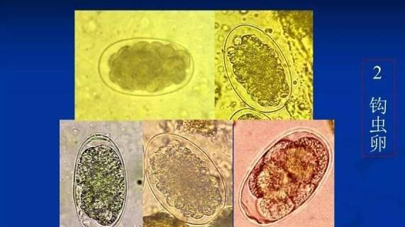
16. Cattle and sheep back-mouth nematode disease:
(1) That is, "hookworm disease", adult parasitism occurs in the small intestines of cattle and sheep.
(2) Morphological characteristics: the head is bent to the dorsal side.
(3) Main route of infection: skin.
(4) Symptoms: diarrhea feces blackening, anemia, emaciation, autumn infection, spring onset.
(5) Diagnosis: fecal examination of insect eggs (dark color, blackening, blunt and rounded at both ends of the eggs, straight on both sides, 8-16 egg cells inside), or autopsy to find the insect body.
(6) Treatment: ambenimidazole, levomimide, ivermectin, mebendazole.
17: Bovine and sheep lung nematode disease:
Parasitizes the lungs of cattle and sheep.
Bovine lung nematode: that is, large lung nematodes, in which the fetal reticulo nematode parasitizes cattle, (the posterior lateral ribs in its croscaria are only separated at the end), of which the posterior lateral ribs are completely fused, in which the filamentous reticule nematode parasitizes sheep, and the posterior lateral ribs in the croscous umbrella are separated only at the end.
Sheep lung nematode: that is, a small lung nematode, mostly parasitic in sheep.
Reservoir: Earthworm.
Symptoms: night and early morning cough, sputum containing insect eggs, difficulty breathing, increased body temperature, emaciation, mostly spring onset.
Fecal detection of eggs: the first stage larvae of filamentous reticulotic nematode stage larvae are blunt and rounded, without button-like protrusions.
Treatment: chloroacetylhydrazide, albendazole, pyrimethazine, ivermectin.
18: Cowhide rope maggot disease:
Caused by the parasitism of the larvae of the striped cord and the cowhide rope in the subcutaneous tissue of the bovine's back.
Form: The rope is large, the body surface is covered with long hairs, and the shape resembles a bee.
Development: Life history belongs to the complete metamorphosis, through the egg, larvae, pupae, rope four stages, the larva by the host skin follicles drilled into the subcutaneous, the entire development period of one year.
Symptoms: The female rope causes strong uneasiness in cattle when laying eggs, and the larvae burrow into the skin, causing pain and itching, especially the tertiary larvae under the skin of the cow's back, causing local connective tissue hyperplasia and subcutaneous cellulitis.
Diagnosis: the larvae appear under the skin of the cow's back, can be palpated to the bulge, have small holes on it, contain the larvae, and squeeze hard to squeeze out the worm body.
Treatment: elimination of larvae in cattle, organophosphorus insecticides, ivermectin.
Often secondary to bacterial infection and suppuration.
19: Sheep Mad Rope Maggot Disease:
Caused by sheep rabid cord larvae parasitizing in the nasal cavity of sheep and its adjacent cavity sinuses, it mainly harms sheep.
Development: It belongs to the complete metamorphosis, and the development passes through 3 stages of larvae, pupae and adult.
Symptoms: commotion of sheep, nostrils against the ground, increased nasal fluid, purulent nasal discharge, wasting.
Diagnosis: spraying liquid into the nasal cavity, collecting nasal ejections, and finding dead larvae.
Treatment: avermectin, dimethoate, cyanatodosamide, with the elimination of the first stage larvae in the nasal cavity as the main measure.