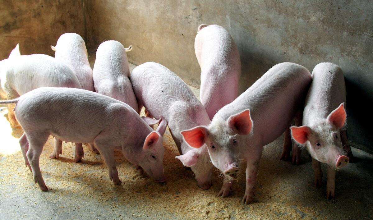
Pig leg crutches: caused by pain, common in 5 causes:
1. Hoof damage (blisters, hoof cracks or deformation);
2. Trauma (stabbing, scratching, bite, tendon or muscle strain, bone damage or misalignment, inappropriate intramuscular injection)
3. Inflammation (streptococcus, parasyllax, mycoplasma);
4. Metabolic disorders (gout);
5. There are five types of allergies (rheumatism).
Principles of treatment of pig leg crutches
Leg crutches: due to pain
Anti-inflammatory + antipyretic analgesic drugs should be symptomatic treatment
Commonly used: cephalosporin, lincomycin, sulfa + salicylic acid and other nonsteroidal antipyretic analgesics
How to differentially diagnose and treat pig leg crutches, the following introduction for reference:
First, leg crutches caused by "hoof damage"
1. Structure of "hoof":
Consists of a shell of dead tissue and living cells that produce keratin, including a very hard outer wall of keratin, a hard base of the hoof, and a softer hoof ball corner (the hoof ball corner surrounds the hoof ball, which relieves the pressure on the hoof, like a mat).
Neither the keratinous crust nor the keratinocytes of the hooves have vascular nerves and cannot feel pain. The dermis layer has blood vessels that supply keratinocyte nutrients and has nerve fibers that can feel pain.
2. Common factors caused by "hoof damage" causing leg crutches:
"Hoof damage" injuries the dermis layer, causing pain due to the pig's own weight pressure and exercise, and is common in:
A. Foot-and-mouth disease
B. Hoof crack or deformation
3. Leg crutches caused by "hoof damage" - foot-and-mouth disease
1) Foot-and-mouth disease
A. It is a highly zoonotic viral and infectious disease
B. It is characterized by the appearance of blisters in the mouth, hooves and milk areas, and the loss of the hoof shell, which induces sudden death of myocarditis.
C. There are 7 serotypes without cross-immunity, including A, O, C, South Africa 1, 2, 3 and Asia 1. Among them: type O mainly causes the onset of disease in pigs, and type I in Asia also causes diseases in cattle and sheep
Foot-and-mouth disease type O also includes: Chinese type (CATHAY type), pan-subtype (ME-SA type), Myanmar 98 lineage (SEA type) Currently: must be immunized to the Myanmar 98 lineage
D. Foot-and-mouth disease vaccine is a "strongly toxic" inactivated vaccine with strong irritation and slow production of antibodies.
The vaccine itself can cause the onset of the disease, so when immunizing, avoid pregnant pigs and nursing sows within 20 days after childbirth, and piglets should be exempted after 50 days of age.
E. Cattle and sheep use foot-and-mouth disease trivalent inactivated seedlings, and some scholars have pointed out that it has positive significance for the prevention and control of foot-and-mouth disease
F. After the occurrence of foot-and-mouth disease, laying mat grass will maximize the survival of the virus
G. Early medication cannot cut off the spread of foot-and-mouth disease, and complete isolation is the correct way.
H. Reducing all stress, especially reducing intramuscular injection, is the main way to reduce death.
I. When there is no specific drug treatment for fever and severe diet reduction, it can be considered:
(1) Banqing granules + VC + amoxicillin Drink water in the whole group to relieve symptoms
(2) Cyclophosphamide, prevent myocarditis
(3) Peracetic acid disinfectant or white vinegar external bath affected part, and then apply the affected part with iodoglycerin, and apply it to the oral cavity with ice boron powder (ice flakes 15 grams, borax 150 grams, glauber's salt 18 grams, a total of fine powder).
4. Leg crutches caused by "hoof damage" - hoof crack or deformation
1) Factors that cause hoof cracks or deformation
A. Seasonal factors:
In autumn and winter, the air turns cool, the capillaries on the surface of the body constrict, and the hooves are poorly flexible and hard and brittle
B. Management:
The ground is too rough (collision), the hooves are pinched or abrasive by gaps in the birth bed, corrosive disinfectants; Inactivity leads to hoof overgrowth (see table) the wall of the trotters of fattening pigs grows 10 mm every four weeks; And for breeding sows over one year old, the wall can still grow 5-6 mm in four weeks) and so on.
C. Variety factors:
Highly selective lean breeds, such as York, Long White, slender limbs and hooves are prone to hair.
D. Nutritional factors:
Biotin, calcium and phosphorus, VD3, zinc, selenium, manganese deficiency and chronic fluorosis. The following nutritional factors caused by hoof damage for your reference:
(1) Biotin:
(VB7, vitamin H, Coenzyme R) Co-A carboxylase co-radical, a rate-limiting enzyme in the first stage of saturated fatty acid synthesis, is also a necessary substance for the synthesis of VC and an important factor for the fixation of CO2 by organisms (carboxylation reaction plays the role of CO2 carrier)
Biotin deficiency, lipid secretion decreased, hoof shell becomes thin and tender, can not maintain the strength and hardness of the hoof cuticle (cuticle is a hydroxyl fatty acid containing 16-18 carbon), hoof keratin will become softer and softer, very easy to wear, resulting in hoof shell cracks, hoof transverse cracks or foot pad cracks.
Factors causing biotin deficiency:
A. The feed energy is too low or the temperature of the pig house is too low, and the energy consumption in the pig body increases.
B. Long-term single use of corn-soybean meal-type diet, lack of biotin-rich raw materials, such as feed yeast, fermentation by-products, etc.
C. Other nutritional factors such as folic acid, pantothenic acid, VB12, VB6, etc. all affect the absorption and metabolism of biotin (synergistic effect of B vitamins)
D, feed contains biotin antagonistic factors, mold, some antibacterial drugs (such as sulfonamides, etc.), oxygen bridge chloroxanaphthalene (insecticide), etc. will cause the loss and inactivation of biotin.
E. Feed oxidation rancidity, hot granulation, improper storage (temperature, humidity) can cause biotin loss
F. The premix is damaged by trace elements such as high copper and choline chloride, resulting in a decrease in the titer of biotin.
G, stress, disease and other factors affecting the function of the digestive tract, so that the biotin synthesis of the large intestine is reduced or the body's absorption is reduced.
(2) Calcium, phosphorus: insufficient content or proportion imbalance of young pigs show rickets, adult pigs form osteochondrosis, in the limbs and hooves show hoof floor fissure and hoof shell keratin softening and bone bending deformation.
(3), VD3: affecting the growth and development of bones, the occurrence of hypochondriasis, limb and hoof irregularities and joint swelling, etc., causing the limbs and hooves of breeding pigs to be unevenly stressed, resulting in cracked hooves, lack of exercise and sun exposure are prone to this disease.
(4) Zinc: it will cause "keratosis imperfecta" (spinous cells are not in the transparent keratinization stage, that is, transformed into the keratinocyte, so that the cells of the stratum corneum contain concentrated nuclei or cell spines that have not disappeared) and lead to hoof side cleft;
(5) Selenium: the main component of glutathione peroxidase, so that the peroxide (H202) (oxidation of the cell lipid membrane and denatured necrosis) is reduced to a non-toxic hydroxyl (-OH) compound.
Denaturation and necrosis of chondrocytes at the end of selenium-deficient bone, muscle atrophy (osteoarthrosis), causing hoof deformation, hair removal, and arthritis.
Glucosamine sulfate can promote the synthesis of cartilage and inhibit the decomposition of articular cartilage
(6) Manganese: mucopolysaccharide polymerase and galactose transferase activator related to chondroitin sulfate in osteogenesis; In addition, manganese only activates alkaline phosphatase, which hydrolyzes pyrophosphate and facilitates bone salt deposition
Disorders of osteogenesis due to manganese deficiency can lead to abnormal deformation of the hooves (short and thick bones, slippage tendons), transverse fissures.
The difference between manganese deficiency and calcium and phosphorus deficiency: normal bone calcification and hard bone, which is different from incomplete bone calcification and bone softness in rickets.
(7) Chronic fluorosis: fluorine can inhibit bone-derived alkaline phosphatase, reduce calcium salt absorption and storage, and cause calcium and phosphorus metabolism disorders. Fluoride is deposited mainly in the teeth (dental fluorosis: white opaque spots and yellow-brown markings) and bones (skeletal fluorosis: joint stiffness, bone deformation) resulting in abnormal deformation and transverse fissure of the hooves.
2) Comprehensive prevention and control of hoof cracks or deformation
A. The cement floor should maintain a suitable smoothness, and the ground should be free of sharp objects and standing water, and it should not be too rough.
B. The pig farm should regularly let the sows go outdoors, exercise their limbs and hooves, and receive sunlight, which is conducive to the synthesis of VD and the absorption of biotin.
C, Wu Kun, etc. believe that in the feed of pregnant and nursing sows (on the basis of using 4% premix), 2% biotin 100g / ton + zinc methionate (organic zinc content ≥ l5%) 300g / ton, continuous use for 14 days, both can treat, but also prevent the disease, and the effect is remarkable.
D. For the affected pig that has just occurred hoof cleft, tie gauze on one end of the wooden stick, and then dip the end of the gauze tied with purple potion, gently pat the hoof, so that the potion can slowly penetrate into the crack, wipe dry, apply erythromycin ointment, once a day, for 5 days.
E. Secondary infection caused by hoof fissure and hoof pad wear, inflamed and swollen limbs, can be used for intramuscular injection of the neck such as penicillin, lincomycin, and sulfonamide.
Second, the leg crutches caused by "trauma"
1. Stabbing injury: deep damage to subcutaneous tissue, external trauma
Features: Ampoule fragments or needles fall inside the circle
Treatment: removal of foreign bodies, without medication, if suppurated, rinsed with hydrogen peroxide.
2. Scratches: superficial damage to skin sharps, external trauma
Features: The height and position of the duckbill drinking fountain are improper, and it is not stopped in time and bites each other in circles
Treatment: length and depth do not cause severe bleeding, without treatment; Severe bleeding requires sutures with spreading penicillin or sterilized crystalline sulfonamides
3. Bite: compound skin damage. External trauma
Features: Stress is greater than the injury itself, and anti-stress is greater than wound management.
Treatment: no bleeding is not treated, bleeding must be disinfected from the wound, at the same time, intramuscular injection of high-dose VC injection.
4. Sprain strain: tendon or muscle strain, internal trauma, sports injury
Features: The circle is wet and slippery, with strong tender points.
Treatment: cold compresses, and anti-inflammatory drugs such as penicillin and cephalosporin.
5. Slip injury: bone damage or dislocation, internal trauma, sports injury
Features: There is a history of parallel bite frame, no tender points in the legs, and edema of one side of the crotch becomes larger.
Treatment: Single-lap feeding or elimination treatment!
6. Inappropriate intramuscular injection: muscle inflammation or necrosis. Internal trauma, mechanical injuries
Features: During the operation of the forward acupuncture point or myotropic acupuncture point, the needle is not sterilized and contaminated or the needle bending damages the muscle tissue or even the needle breaks in the muscle, causing inflammatory edema of the muscle or the formation of pustules.
Treatment: leave the pus untreated for the time being, wait for the pus to mature, cut and drain the pus, and rinse with hydrogen peroxide.
Third, the leg crutches caused by "inflammation"
1. Arthritis streptococcus: (Gram-positive bacteria)
Features: Deformed bones or suppuration – with pimples
Infectious, soft or hard, caused by the E-group serotype, (16 serotypes in total)
Treatment: Gram-positive bacteria, preferably lincomycin or sulfonamides.
2. Parasynine (Glazer's disease) :( Gram-negative bacteria)
Features: Water in the joint cavity - walking legs are stiff, goose-shaped steps will not bend
When the water is not serious, the appearance is normal; If there is too much water, the joint membrane ruptures, the water leaks, the whole leg is swollen;
Treatment: Gram-negative bacteria, preferred quinolones (sathorins) and aminoglycosides (amikacin)
3. Mycoplasma synovitis: characteristics of acute claudication behavior
Mycoplasma: Gram-negative, the only prokaryotic cells without a cell wall, organelles with only ribosomes
It has a strong affinity for the thymus, peritoneum, interstitial cells of the joint synovial cyst membrane and the central nervous system
The protective period is at least 60 weeks after the onset of mycoplasmic pneumonia in pigs and 12 to 30 months after infectious pleural pneumonia in cattle.
After mycoid infection, the body mainly causes humoral immune responses, which can produce IgM, IgG, and IgA.
Features: Acute claudication behavior, causing acute synovitis and arthritis in young pigs, present in the joints, mucosal secretions and lymph nodes in the acute claudication phase.
Treatment: resistance to penicillin, mucobacterin, sensitivity to doxycycline, Tylosin, Linco
Fourth, the leg crutches caused by "metabolic disorders"
Gout: refers to a metabolic disease that occurs when the feed contains inferior meat meal, fishmeal and other animal protein raw materials rich in purine and alkali, and contains too much calcium and magnesium, as well as long-term va, or renal insufficiency (drugs), resulting in abnormal purine metabolism resulting in increased uric acid synthesis or decreased uric acid clearance.
Uric acid in the blood plasma reaches saturation, causing uric acid monosodium crystals to deposit in relatively vascular-deficient tissues around the distal joint. This crystallization can lead to acute inflammatory synovitis in single or multiple joints.
Features: slow movement, joints slightly swollen due to urate deposition, initial soft and painful after the swelling of the part gradually hardened to form nodules, nodules rupture and discharge gray-yellow cheese-like.
treat:
A. Acute phase: high-dose nonsteroidal anti-inflammatory drugs (angustin) + colchicine
B. Remission period: uric acid-lowering therapy (including allopurinol that inhibits uric acid production and atholfon or benzbromomarone that promotes uric acid excretion)
Fifth, "allergic reactions" caused by leg crutches
1, rheumatism: (customary rheumatism, specifically refers to "rheumatic fever"), in fact, "rheumatism" is not a specific disease, but a general term for a type of disease.
It is a group of diseases that invade joints, bones, muscles, blood vessels and related soft tissues or connective tissues, most of which are autoimmune diseases, most of which have genetic predispositions, and different autoantibodies can be detected in the blood.
Autoimmune diseases: under the inducement of antigen factors (hidden antigen release (such as special anatomical positions such as lens and sperm of the eye) and autoantigen changes (such as rheumatoid factor)), genetic factors, immune factors, etc., autoimmune tolerance is broken, and the immune response continues to be prolonged, resulting in self-cell destruction, tissue damage or abnormal function.
Immune tolerance: the normal body's immune system is in a phenomenon of no response or weak response to autoantigens (the fetal immune system in the embryonic stage can only be exposed to autoantigens, so it is immune to autoantigens after birth).
1) Rheumatic mechanism:
Suppressive T cells are lowly functioning, humoral immunity is hyperfunction, antigens stimulate the activation of corresponding T cells, and some T cells produce a variety of inflammatory cytokines; Some T cells reactivate B cells to produce antibodies, form an immune complex with antigens, and deposit them in tissues to cause disease.
2) Rheumatism characteristics: fast erythrocyte sedimentation rate (anticoagulant blood into the erythrocyte tube vertically standing, red blood cells sink due to large density. Usually the distance at which red blood cells sink at the end of the first hour indicates the sedimentation rate of red blood cells), often with autoantibodies, migratory polyarthritis, no definite tender points, and forced exercise relief.
3) Rheumatic classification:
1. Diffuse connective tissue disease: characterized by loose connective tissue myxoid edema and fibrin degeneration
A. Rheumatoid arthritis: small joints, symmetrical joint swelling and pain, multi-organ damage
B. Systemic lupus erythematosus: mucosal ulcers, alopecia, discoid erythema, arthritis and deformities, and multiple organ damage
C. Scleroderma: the skin thickens and hardens, so the appearance is tight and waxy
D. Primary Sjogren's syndrome: salivary glands and tear glands are damaged and dry mouth and eyes, rampant caries, light sensitivity
E. Dermatomyositis: lymphocyte infiltration of striated muscles (skeletal muscles, myocardium), characterized by symmetrical limb band muscle, neck muscle, pharyngeal muscle weakness, and difficulty in looking up and swallowing.
F. Vasculitis (polyarteritis nodosa): subcutaneous nodules distributed along the superficial arteries (more likely to occur at arterial bifurcations, showing segmental features), reticular blue spots, unexplained testicular pain, lower extremity tenderness.
2. Spinal arthropathy: blood rheumatoid factor negative, interrelated multisystem chronic inflammatory rheumatism.
Clinically seen diseases include:
A. Forced spondylitis: low back pain, aggravated at rest, accompanied by heel pain, red eye
B. Psoriasis (psoriasis) arthritis:
There is a psoriatic rash (inflammatory red halo around the erythema, the surface is covered with multiple layers of silver-white scales, easy to scrape off), lytic bone lesions at the end of the fingers, x-rays are "pen cap-like" changes, and the erythrocyte sedimentation rate is normal.
3. Rheumatism associated with infection: microorganisms produce antigens or superantigens to stimulate or initiate an immune response.
1) Rheumatic fever:
Hyaluronic acid of the capsules of group A hemolytic streptococcal has a common antigenicity with hyaluronic acid of synovial membrane and joint fluid, which avoids leukocyte phagocytosis. It shows multiple, wandering, and multiple violations of large joints, closely related to weather changes, salicylic acid is effective, arthritis disappears with rheumatism, and no rigidity or deformities are left.
B. Reactive arthritis:
A variety of bacteria, viruses, bacteroids and other pathogens cause urethral and intestinal infections 1-6 weeks after the occurrence of asymmetric acute non-septic arthritis, and the joints are obviously swollen and form dachshund fingers.
2) Comprehensive prevention and treatment of rheumatism:
A. Non-steroidal antipyretic analgesics such as salicylic acid
B, dimi and other corticosteroid drugs:
C. Slow-acting anti-rheumatic drugs: such as: sulfasalazine (oral does not absorb sulfonamides, inhibits the synthesis of immune complexes and rheumatoid factors, and is also used for the treatment of ulcerative colitis)
D. Immunosuppressants: methotrexate (antifolate antineoplastic drugs, inhibit dihydrofolate reductase, so that dihydrofolate can not be reduced to biologically active tetrahydrofolate, so that the metastatic effect of a carbon group in the biosynthesis process of purine and pyrimidine is blocked, and the synthesis of tumor cells is hindered; Treatment of rheumatoid in combination with adalimumab can slow the progression of joint injury); Azathioprine (an imidazole derivative of 6-mercaptopurine, can produce an alkylation effect to inhibit the synthesis of accounting and prevent cell proliferation. antimetabolites with an immunosuppressive effect for the treatment of systemic lupus erythematosus); Cyclophosphamide (an antitumor drug, which is converted into unstable aldehyde phosphoramide by microsomal functional oxidase in the liver and decomposes into amide chlorambus and acrolein in tumor cells, the former has cytotoxic effect on tumor cells, cross-links with DNA, and inhibits DNA synthesis). (Source: Dr. Kang)