The ligaments around the ankle can be divided into 3 groups depending on their anatomical location:
Lateral collateral ligament
Medial collateral ligament (triangular ligament)
Inferior tibia-fibula joint ligament
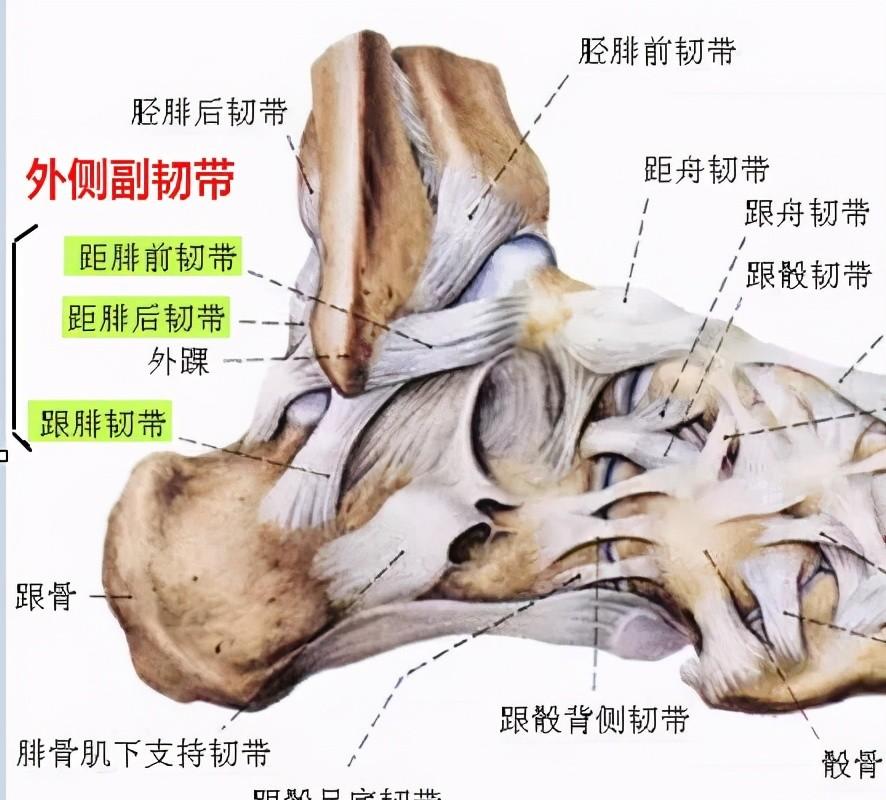
The lateral collateral ligaments
Lateral collateral ligament complexes include: anterior ligament from the fibula, achillesifular ligament, and posterior ligament from the fibula.
Anterior talofibular ligament: from the anterior edge of the lateral malleolus, obliquely descending anteriorly to the lateral side of the tal neck, 2 to 2.5 mm thick. In the neutral position, it is parallel to the long axis of the foot from the anterior fibula ligament and perpendicular to the long axis of the calf. The main effect is to limit the anterior movement of the talus.
Calcaneofibular ligament: Starts from the tip of the lateral ankle and slopes backwards to the lateral side of the calcaneum, deep in the tendons of the long and short tendons of the fibula. The main effect is to limit calcaneal varus.
Posterior talofibular ligament: The posterior ankle fossa from the posterior part of the lateral malleolus and ends horizontally in the posterolateral process of the tal bone, making it the strongest of the 3 bundles. The main effect is to limit the talus backwards.
Ankle lateral ligament injuries: very common, with the highest number of varus sprains, accounting for 85%. Of the sports injuries of basketball players, 53% are those of the lateral collateral ligament of the ankle joint, and 21% of the sports injuries of football players are injuries of the lateral collateral ligament of the ankle joint.

For the convenience of treatment, ankle lateral ligament injuries are classified as follows:
I. Ligament strain, no tearing, stable joints, no damage to function;
II. Injury to the heel-fibula ligament or anterior fibula ligament, moderate pain, swelling, joint instability;
III. Simultaneous injury of the heelofiliary ligament and the anterior fibula ligament, pain, swelling, and joint instability.
↓ Distance from the anterior fibula ligament: normal and tear
<h1 class="pgc-h-arrow-right" data-track="31">deltoid ligament</h1>
The triangular ligament works with other structures of the ankle to maintain ankle stability. The triangular ligament is a composite ligament with a fan-shaped structure consisting of shallow and deep layers.
Includes: tibial pitch ligament (anterior/posterior), tibia heel ligament, shin boat ligament.
↓ Shin boat ligament
↓ Tibi ligament
↓ Tibial anterior ligament
↓ Posterior tibial ligament
↓ Posterior tibial ligament injury
The distal joint surface of the tibia-fibula consists of a convex surface at the distal tip of the fibula and a concave surface at the distal tibia, linked by the tibia-fibular ligament, without cartilage structures within the joint. The distal tibia-fibula combined ligament consists of four-part ligaments, including: interosseous ligament, anterior tibia-fibular ligament, posterior tibial-fibula ligament, and inferior transverse ligament.
The interosseous ligament is formed by thickening the distal end of the interosseous membrane. The anterior tibial-fibular ligament begins on the anterolateral side of the tibia nodule and ends on the anterior side of the fibula ridge. The posterior ligament of the lower tibia-fibula begins on the posterior side of the tibial spine and ends at the posterior edge of the lateral malleolus. The inferior transverse ligament forms the lower half of the posterior ligament of the posterior tibial fibula and can be considered part of the posterior ligament of the posterior tibial fibula.
Posterior tibial-fibula ligament (35%), deep posterior tibial-fila ligament (33%) had the greatest effect on ankle stability, followed by interosseous membrane (22%) and superficial posterior tibial-fibular ligament (9%).
The mechanisms of injury to the combined ligament of the lower tibia-fibula are usually ankle external rotation and excessive dorsal flexion. Sports that can cause joint injuries to the lower tibia include sports (soccer, etc.), low-energy trauma, etc. External spin injuries usually occur in the anterior or posterior position of the ankle. Tibia-fibula joint ligament injuries can occur solitaryly or with fractures.
Tibial-fibula combined with distal ligament node structures, including: interosseous ligament (IOL), inferior tibia-fibula anterior ligament (AITFL), posterior tibial-fibula ligament (PITFL), inferior transverse ligament (ITL).
Three imaging tests can be used to diagnose combined ankle tibia-fibula injury: tibiofibular overlap distance, tibiofibular clear space, medial joint space, etc.
Measurement of tibial-fibula overlap greater than 6 mm above the tibial fornix fornix at 1 cm above the tibial fornix fornix, or greater than 1 mm overlap of ankle acupuncture point x-rays indicates normal, and inferior tibia-fibula joint injury if the anteroposterior x-ray overlap is less than 6 mm.
The width of the medial space of the ankle should be equal or slightly smaller than the tibial vault/talparagus gap.
Decreased tibia-fibula overlap distance on load-bearing or non-load-bearing X-rays, and increased tibial-fibular space or medial space of ankle acupuncture suggest joint tibia-fibula injury.
The tibia-fibular space is the most reliable measure of imaging, as it is rarely affected by the angle of the lower extremities and radiography. CT can detect fractures that are not obvious on X-rays, while MRI is extremely sensitive and specific for diagnosing tibia-fibula joint ligament injury.
Tibial-fibular combined injury diagnosis measurement lines: A, tibia-fibula overlap, B, tibia-fibular space, C, medial space. (Where the tibia-fibula overlap and the tibia-fibular space are measured at a distance of 1 cm from the tibia dome.) )
(The above content is compiled from the Network)